Simple Method Developed to Characterize Immune Cells in Tumors
By LabMedica International staff writers
Posted on 27 Jul 2016
Despite recent achievements in the development of cancer immunotherapies, only a small group of patients typically respond to them and therefore predictive markers of disease course and response to immunotherapy are urgently needed.Posted on 27 Jul 2016
A new method has been developed for analyzing multiple tissue markers using only one slide of a tumor section to better understand immune response occurring locally. The multiplexed immunohistochemical consecutive staining on a single slide (MICSSS) helps characterize human cells involved in immune responses at the tissue site, before and after treatment with immunotherapy.

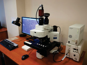
Image: A whole-slide scanner with OlyVIA software (Photo courtesy of Olympus).
Scientists at the Icahn School of Medicine at Mount Sinai (New York, NY, USA) and their international colleagues obtained paraffin-embedded human tonsils, ulcerative colitis, non-small cell lung cancer (NSCLC), melanoma, and colorectal tumor samples from their Biorepository tissue bank. The formalin-fixed paraffin embedded (FFPE) were processed for immunohistochemistry (IHC) and images were acquired using an Olympus whole-slide scanner with OlyVIA software (Olympus Life Science, Center Valley, PA, USA) or an Eclipse Ci-E microscope (Nikon Instruments, Melville, NY, USA).
The authors have described a multiplexed chromogenic IHC strategy for high-dimensional tissue analysis that circumvents many of the limitations of regular chromogenic, immunofluorescence, and mass cytometry approaches that could be readily implemented in clinical pathology laboratories. The MICSSS method provides a new powerful tool to map the microenvironment of tissue lesional sites with excellent resolution, in a sample-sparing manner, to monitor immune changes in situ during therapy and help identify prognostic and predictive markers of clinical outcome in patients with cancer and inflammatory diseases.
Sacha Gnjatic, PhD, an Associate Professor and senior co-author said, “Our goal was to get a better understanding of immunologic responses at the tumor site while addressing the need for high-dimensional analysis using as little tissue as possible. We need more comprehensive analyses of the immune microenvironment of tumors, as part of our immune monitoring to inform treatment and predict outcomes for cancer patients.” The study was published on July 14, 2016, in the journal Science Immunology.
Related Links:
Icahn School of Medicine at Mount Sinai
Olympus Life Science
Nikon Instruments


