


Researchers Evaluate Automated Tissue Analysis Across Four Cancer Types
Posted on 20 Nov 2026
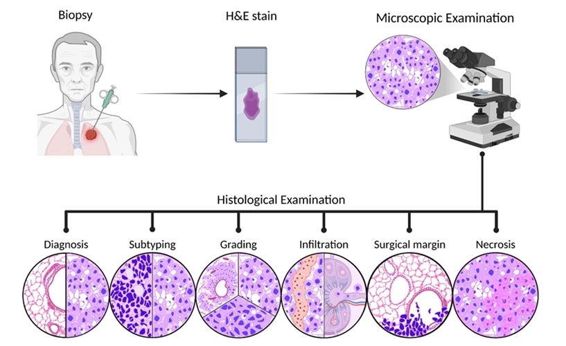
Histopathology image analysis plays a central role in cancer diagnosis, but tumor tissue can vary considerably across different regions of the same specimen. Pathologists examine cellular morphology, nuclear features, tumor–immune cell interactions, and tumor boundaries, which can differ by cancer type and clinical context. This variability presents challenges for consistent and reproducible image interpretation. New research evaluates the performance of computer vision tools in analyzing tumor tissue images across multiple cancer types.
Researchers at the Skolkovo Institute of Science and Technology (Skoltech), in collaboration with the University of Sharjah in the United Arab Emirates, Tokai University in Japan, and other institutions, evaluated computer vision techniques for histopathology analysis across lung, breast, colorectal, and prostate cancers. The review examined how algorithms identify tissue characteristics relevant to different diagnostic tasks. In lung cancer, these methods can support tumor subtype classification based on tissue structure, while in breast cancer, they can help assess tissue morphology and receptor marker proteins in specially stained specimens.
The researchers compared five computer vision tools using publicly available histopathology images representing four cancer types. Performance was assessed by comparing automatically identified cell nuclei with expert-annotated reference images. Cellpose and StarDist, implemented through Fiji, used deep learning-based segmentation, while CellProfiler and Icy relied on pixel intensity thresholds in this evaluation. Ilastik was trained using a limited number of annotated examples but sometimes merged neighboring nuclei, highlighting differences in how the tools handled complex tissue structures.
Cellpose achieved the highest mean Dice score in four of six comparisons, including the pooled MoNuSAC evaluation. The Dice score measures agreement between automated segmentation and expert annotations, with a value of 1 indicating complete agreement. In colorectal cancer images, Cellpose and StarDist achieved similar performance, both scoring 0.74. However, StarDist performed slightly better in low-cell-density triple-negative breast cancer images, achieving a mean Dice score of 0.84 compared with 0.82 for Cellpose. The researchers also observed that StarDist occasionally missed nuclei in densely packed tissue, demonstrating how tissue characteristics can influence segmentation accuracy.
Published in Discover Artificial Intelligence, the review highlights the potential of computer vision to support histopathology analysis while emphasizing the need for further clinical validation. The authors noted that broader evaluations across multiple institutions are necessary to establish reliability in routine practice and determine how AI-assisted analysis influences pathologists’ decisions. They also stressed the importance of interpretability, allowing pathologists to identify which regions of a tissue slide contributed to an algorithm’s findings.
“You need to start not with an algorithm, but with the question that the doctor will have to answer based on the histological specimen,” said Svetlana Illarionova, the review's first author, assistant professor and head of research group at the Skoltech AI Center.
“In some cases, it's important not to miss a tiny focus; in others, it's important to see exactly how different cells are arranged in the section. And in any case, the doctor must see which areas of the image the model is based on. Its reliability should be tested using materials from different laboratories, where they are stained and scanned in different ways,” added Illarionova.
Related Links
Skoltech
University of Sharjah
Tokai University






.jpg)