Rapid Mass Spectrometry Test May Aid Glioma Margin Decisions
Posted on 05 Oct 2026
Glioma brain tumors are highly infiltrative and can extend into nearby healthy brain tissue, making tumor margins difficult to define during surgery. Residual tumor cells may contribute to recurrence and complicate subsequent treatment, while standard molecular methods generally provide results only after the procedure. To support faster intraoperative assessment, researchers have developed a mass spectrometry method that can evaluate resected brain tissue in about three minutes.
Purdue University (West Lafayette, IN, USA) researchers, working with Mayo Clinic (Rochester, MN, USA), developed a syringe touch spray approach for rapid molecular assessment of glioma tissue. The method uses ambient ionization mass spectrometry to analyze endogenous compounds from brain tissue in an open environment. In the workflow, fractions of samples taken during brain tumor surgery are transferred onto the plunger of a hypodermic needle, sprayed with a solution, and analyzed by tandem mass spectrometry.
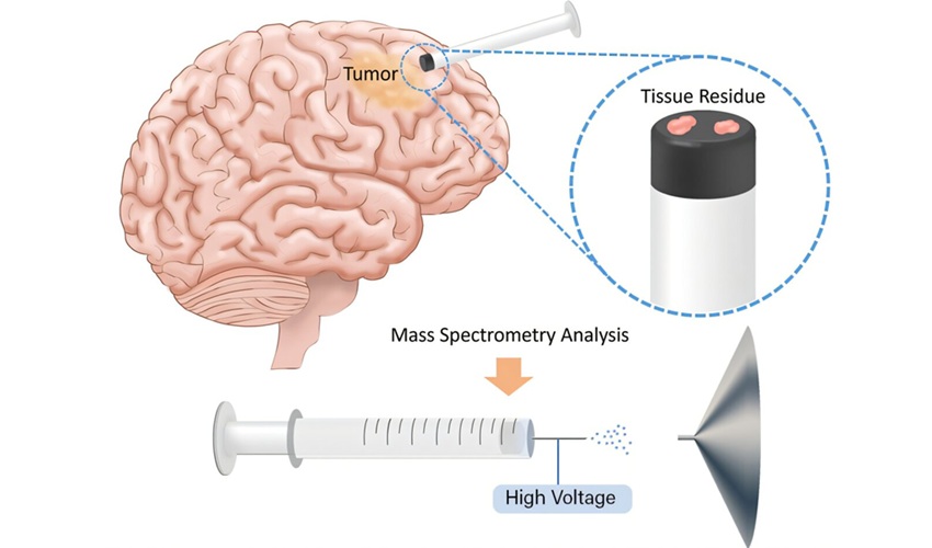
The study used 86 human brain samples obtained through MRI-guided precision surgery. Researchers measured ratios of signals for multiple pairs of endogenous brain compounds. These metabolite ratios distinguished glioma from nontumor tissue with high confidence and also provided information on tumor subtype, grade, and mutation status.
The work was published in Proceedings of the National Academy of Sciences on September 30, 2026. The collaborating institutions were Purdue University and Mayo Clinic. The team reported that further evaluation would be needed during surgery, including a similar study involving more than 50 patients, before a clinical trial could be proposed.
“This technology would not replace the current standard of care during brain tumor surgery. Instead, it could provide surgeons with molecular information that is not currently available in the operating room, helping to guide surgical decisions during tumor removal, particularly at the tumor margins,” said R. Graham Cooks, Henry Bohn Hass Distinguished Professor of Chemistry at Purdue University.
Related Links
Purdue University