Handheld Device Improves Targeted Cell Collection for Early Ovarian Cancer Assessment
Posted on 27 Sep 2026
High-grade serous ovarian cancer is the most common form of ovarian cancer, and many cases are thought to originate in the fallopian tubes. Because precursor lesions can be microscopic, they are difficult to sample and detect at an early stage. Five-year survival can exceed 90% when ovarian cancer is diagnosed early, but falls below 50% when the disease is detected at stages 3 or 4. To improve access to these hard-to-sample tissues, researchers have developed a device that collects living cells from specific anatomical sites.
Researchers at the Massachusetts Institute of Technology (MIT; Cambridge, MA, USA), working with colleagues at Johns Hopkins University (Baltimore, MD, USA), developed a handheld fluidic-shear device for targeted cell collection. The 3D-printed microfluidic device uses one syringe to create a vacuum seal against the tissue. A second syringe drives liquid through a channel, generating shear stress that gently detaches cells from a small area while leaving surrounding tissue intact.
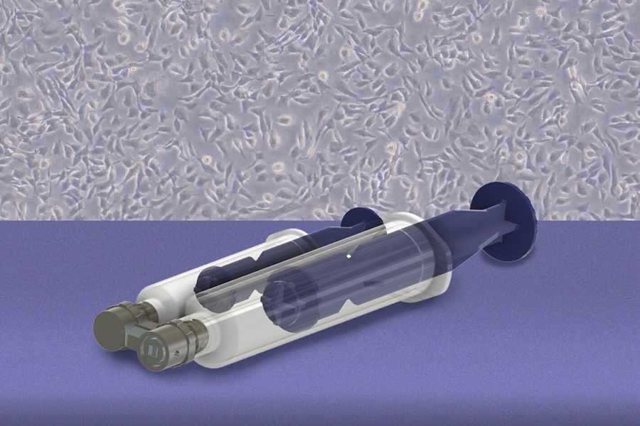
In a study published in Device on September 24, 2026, the researchers used the system to sample newly excised tissue and recover living cells for cultivation. They tested it on fresh human fallopian tube samples and compared its cell-collection performance with conventional approaches. Cells collected with the device remained viable and grew in culture more readily than cells detached by conventional methods. The team also used collected fallopian tube cells to grow organoids before returning the tissue samples for conventional pathology.
Tests across different cell types showed that the applied shear stress could be adjusted for collection. Loosely adherent prostate cancer cells detached at 1 pascal of stress. By contrast, only a few bone cancer cells detached at 5 pascals.
The researchers expect initial use to focus on tissue already removed from the body. They are also exploring optical approaches to identify suspicious regions that could then be sampled with the device. Future goals include collecting cells inside patients and using living patient-derived cells to study disease and test treatment responses.
“We wanted to collect living cells from specific regions of the fallopian tube while leaving the surrounding tissue intact. Once we have these living cells, there are many things we can do with them. We can use them for diagnostics, grow them into organoids and build living models of disease. Ultimately, this could allow us to test how an individual patient's cells respond to different treatments and help us develop more personalized medicines,” said Kripa Varanasi, senior author of the study and professor of mechanical engineering at MIT.
Related Links
MIT