Noninvasive Imaging Approach Aims to Detect Basal Cell Carcinoma Before It Becomes Visible
Posted on 23 Sep 2026
Basal cell carcinoma is the most common form of skin cancer and can damage nearby structures such as the nose or eyes when it develops on the face. Diagnosis typically depends on visible skin changes, which can limit opportunities for earlier, less invasive treatment. A new feasibility study now shows that combining line-field confocal optical coherence tomography with artificial intelligence may reveal tumors before they become clinically apparent.
At Friedrich-Alexander-Universität Erlangen-Nürnberg (FAU), investigators developed a systematic facial screening workflow called SUBSCANS that integrates line-field confocal optical coherence tomography (LC-OCT) with AI-assisted image recognition. LC-OCT combines optical coherence tomography (OCT), which indicates tumor thickness and extent, with confocal microscopy to capture cellular-level detail. The system generates vertical, horizontal, and three-dimensional images of skin in real time at micrometer-scale resolution, and the AI overlays a color-coded probability value for basal cell carcinoma while leaving the final diagnosis to the physician.
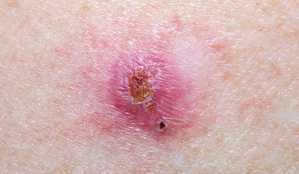
In the feasibility study, clinicians systematically scanned the faces of patients at high risk for skin cancer. Using this procedure, they detected basal cell carcinoma before any visible changes to the skin developed for the first time. LC-OCT is already used as standard care to identify early basal cell carcinoma and to monitor the success of noninvasive therapies, and SUBSCANS is intended to shift detection even earlier in high-risk populations.
The work was conducted by the Department of Dermatology at Uniklinikum Erlangen and was published in JAMA Dermatology. The team notes that routine use of SUBSCANS is not yet possible, citing a current lack of sensitivity data and the time required to perform the procedure.
“Detecting basal cell carcinoma early allows for less invasive treatment options. In some cases, all that is required is a cream,” said Dr. Moritz Ronicke, Department of Dermatology at Uniklinikum Erlangen.
“Our new approach using ‘SUBSCANS’ would allow patients at a high risk of developing skin cancer to receive a diagnosis even earlier on. If the SUBSCAN is routinely used in future, this could lead to more widespread use of creams for treating basal cell carcinoma, even avoiding the need for surgery in such cases,” added Dr. Ronicke.
Related Links:
FAU