AI Spatial Analysis of Routine Slides Helps Predict Pancreatic Cancer Recurrence
Posted on 11 Sep 2026
Predicting which patients with pancreatic cancer will recur after treatment and surgery remains challenging. Pathology evaluation typically measures residual tumor burden, but recent work found that tumor amount alone did not reliably distinguish higher- from lower-risk patients. The spatial organization of residual cancer within surrounding stroma may add prognostic value, especially when neoadjuvant chemotherapy produces only a limited pathologic response. A new study shows that AI-enabled spatial analysis of routine hematoxylin and eosin (H&E) slides can help identify patients at higher recurrence risk.
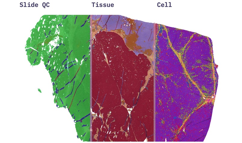
Mayo Clinic (Rochester, MN, USA) researchers developed an AI-enabled spatial analysis approach that reads organizational features on standard pathology slides. The method uses an AI tool to identify cancer and stromal regions, then quantifies how those regions form patches, boundaries, and intermixed areas. Investigators adapted techniques from landscape ecology to measure tissue shape, fragmentation, and the degree of cancer-stroma intermixing on H&E slides.

The study analyzed tissue from 203 patients with pancreatic ductal adenocarcinoma who received treatment before surgery but showed only a limited pathologic response. Two spatial signatures predicted disease-free survival even after accounting for stage, lymph node status, and other established clinical and pathologic risk factors. Patients whose residual cancer showed a more fragmented, intermixed pattern with surrounding stroma experienced earlier recurrence, whereas residual tumor amount alone did not reliably stratify risk.
Across models, the AI-derived spatial features improved separation of higher- and lower-risk groups when conventional measures were less informative. In one model, patients classified as high risk had a 71% higher adjusted risk of recurrence; in another, their adjusted risk was more than doubled. These high-risk spatial patterns also showed fewer immune cells within cancer regions, with immune cells tending to cluster around the tumor rather than infiltrate it.
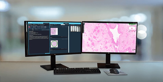
Because the workflow analyzes slides already generated during routine care, it may provide added prognostic information without requiring another tissue test. The study was published in Clinical Cancer Research, and the researchers note that prospective studies are needed before the approach can inform clinical decision-making.
"Current pathology assessments largely tell us how much tumor is left after treatment. We wanted to know whether the geography of that remaining cancer could reveal additional biology about recurrence risk," said Ryan Carr, M.D., Ph.D., a Mayo Clinic oncologist and senior author of the study.
"Our long-term goal is to better identify which patients remain at greatest risk and ultimately use that knowledge to guide more individualized surveillance, adjuvant therapy and clinical trial design," added Carr.
Related Links
Mayo Clinic