Label-Free Imaging Reveals Hidden Immune Activity in Blood Samples
Posted on 03 Sep 2026
Peripheral blood mononuclear cells (PBMCs) are widely used to monitor infection, autoimmunity, cancer, and treatment response, yet most assays depend on fluorescent labels that can perturb cells and provide limited functional insight. Measuring immune-cell metabolism nondestructively has remained a diagnostic challenge. Functional readouts at single-cell resolution could sharpen interpretation of immune dynamics in blood. New findings demonstrate a label-free optical approach that gauges immune-cell metabolic activity directly within mixed PBMC samples.
At the Morgridge Institute for Research (Madison, WI, USA), investigators used optical metabolic imaging (OMI) to interrogate immune-cell function without extrinsic dyes. The method excites endogenous metabolic cofactors with two-photon microscopy and measures the time these molecules emit light, generating fluorescence lifetime signatures that correlate with cellular activation state. Because OMI relies on native signals, analyzed cells remain viable for subsequent testing.
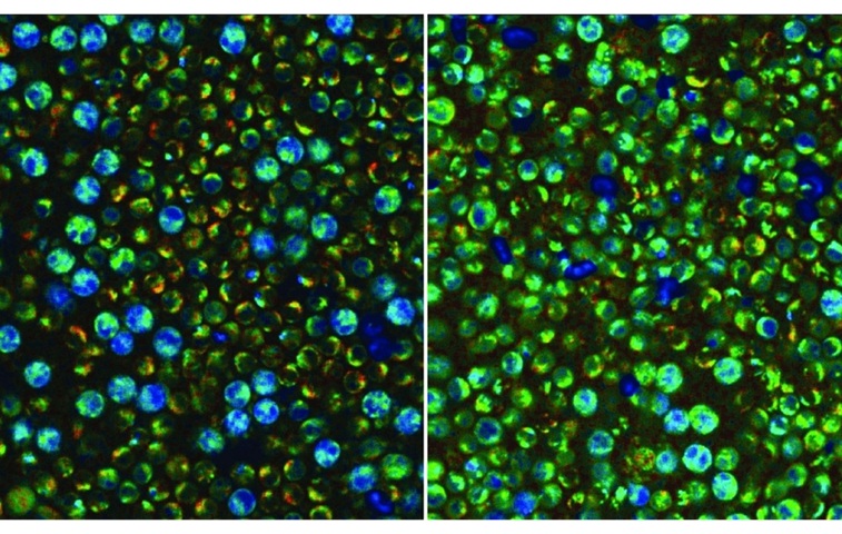
The study examined PBMCs isolated from three healthy donors and analyzed thousands of individual cells in resting and activated states. Researchers applied machine-learning algorithms to determine whether metabolic measurements alone could identify immune-cell subtypes and detect early activation within heterogeneous blood samples. The single-cell approach captured metabolic heterogeneity that would be obscured by bulk measurements.
OMI distinguished activated from resting PBMCs with nearly 94% accuracy just two hours after stimulation. In resting samples, monocytes were identified with 96% accuracy and with 88% accuracy after activation, while natural killer (NK) cells were identified with approximately 74% accuracy in both states. By contrast, T cells and B cells displayed more similar metabolic profiles under the experimental conditions, reflecting less distinctive signatures in this framework.
According to the release, the technique is currently a research tool and does not yet match the accuracy of established labeling methods for identifying every immune-cell subtype. The work, published in Biophotonics Discovery on September 1, 2026, also notes opportunities to enhance deployment by pairing advanced biophotonic imaging with machine learning for broader studies of immune function in health and disease.
Related Links
Morgridge Institute for Research
University of Wisconsin–Madison





.jpg)