New Tissue Mapping Approach Identifies High-Risk Form of Diabetic Kidney Disease
Posted on 01 May 2026
Diabetic kidney disease is a leading cause of chronic kidney disease and end-stage kidney disease, affecting 20%–40% of people with diabetes and more than 107 million individuals worldwide as of 2021. Despite routine monitoring with kidney function tests and proteinuria, clinicians often cannot explain the wide variability in disease progression among patients. New findings offer further insight, identifying a distinct B cell–rich intrarenal phenotype linked to faster progression through high-resolution spatial mapping of human kidney tissue.
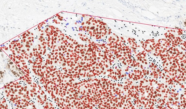
Researchers at the Perelman School of Medicine at the University of Pennsylvania (Philadelphia, PA, USA) created a detailed spatial map that preserved tissue architecture while measuring gene activity directly in kidney samples. The approach identified which cells were present, where they were located, and how they interacted. Using an unbiased, near–genome-wide spatial method, the team characterized disease features at cellular resolution.
")
Kidney tissue from dozens of patients was examined, encompassing more than five million cells. The analysis revealed recurrent patterns of cell groupings and interactions, including a scarring- and inflammation-associated tissue pattern that increased with worsening disease. These data formed an atlas of how diabetic kidney disease remodels the organ.
Within inflamed regions, some patients exhibited organized B-cell clusters resembling immune structures typically seen in autoimmune diseases. Patients with these B cell–rich regions experienced markedly faster progression to kidney failure, and the same areas contained other immune cells that support B cells. The investigators then derived a gene-based signature and a blood test to help identify this high-risk subgroup without requiring comprehensive tissue mapping.
The study, published in Nature on April 29, 2026, indicates that diabetic kidney disease comprises biologically distinct subtypes and highlights the value of interrogating disease directly in intact tissue. The authors note that understanding intrarenal inflammatory organization may inform more targeted therapeutic choices and that spatially resolved, near–genome-wide analyses can reveal patterns missed in dissociated cell studies.
“Diabetic kidney disease has often been treated as a single condition, but patients can have very different outcomes. By looking directly at the kidney tissue, we can now see different disease processes and start to match treatments to what's actually happening in each patient,” said Katalin Susztak, MD, Ph.D., a professor of Renal Electrolyte and Hypertension and co-director of the Penn/CHOP Kidney Innovation Center at the Perelman School of Medicine at the University of Pennsylvania.
“Understanding how inflammation is organized within the kidney gives us a new way to classify disease. This could lead to more precise treatments tailored to each patient,” said Bernhard Dumoulin, MD, a postdoctoral fellow in the Susztak lab and first author of the study.
Related Links
Perelman School of Medicine at the University of Pennsylvania