High-Accuracy Tumor Detection Method Offers Real-Time Surgical Guidance
Posted on 15 Oct 2025
Pancreatic neuroendocrine neoplasms (PNENs) are rare cancers that affect hormone-producing cells in the pancreas. Although uncommon, their incidence has been increasing, and surgery remains the only curative option. However, surgical success often depends on pathology results that can take hours or days, delaying decisions and raising the risk of incomplete tumor removal. Now, a new imaging approach promises to provide real-time detection of these tumors with high precision.
Researchers at the University of Arizona (Tucson, AZ, USA) have developed a powerful imaging and analysis method that combines label-free multiphoton microscopy (MPM) with deep learning. The technique uses light-based imaging to capture natural fluorescence in tissue without staining or damaging samples. This allows clear visualization of tumor structures during surgery, enabling faster and more accurate decision-making for cancer treatment.
")
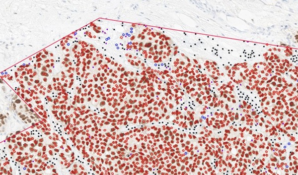
In the study, published in Biophotonics Discovery, the team used MPM to scan pancreatic tissue samples for natural fluorescent markers such as collagen, NADH, FAD, lipofuscins, and porphyrins. These biomarkers help distinguish between healthy and cancerous tissue. To classify the images, researchers trained one machine learning (ML) algorithm and four convolutional neural networks (CNNs), teaching them to recognize subtle differences between tumor and normal tissues.
The CNNs achieved classification accuracies between 90.8% and 96.4%, outperforming the traditional ML model, which reached 80.6% accuracy. The models were tested using samples from multiple biorepositories, demonstrating robustness across varied sources. Analysis of the ML algorithm revealed that collagen levels and image contrast were key indicators of cancer, offering biological insights into PNEN tissue characteristics.
In addition to the high accuracy, the study found that MPM imaging is considerably faster than conventional histology and could be adapted for intraoperative use. Future work will focus on testing the technique with fresh tissue during surgery and expanding its capabilities to determine tumor grade and type. Such advancements could make real-time, automated digital pathology a clinical reality.
This integration of optical imaging and artificial intelligence represents a step forward in precision oncology, potentially reducing repeat surgeries and improving surgical outcomes for pancreatic cancer patients.
Related Links:
University of Arizona