New AI Tool Outperforms Previous Methods for Identifying Colorectal Cancer from Tissue Sample Analysis
Posted on 03 Mar 2025
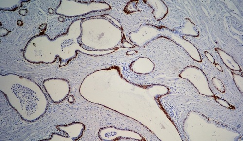
Tissue analysis typically involves a pathologist reviewing scanned digital slides from a patient’s intestinal sample and marking specific areas, such as those where cancerous and related tissues are present. A new artificial intelligence (AI)-driven tool has now been developed to identify colorectal cancer in tissue samples that surpasses all previous models in terms of classification accuracy.
This tool was the result of a collaborative study led by researchers at the University of Jyväskylä (Jyväskylä, Finland). The AI model created in this study demonstrated superior performance in classifying tissue samples compared to prior models. This new tool has the potential to streamline the process for doctors by automating the analysis of tissue slides. It analyzes samples and highlights areas with various tissue types, offering a level of accuracy that could greatly reduce the workload of histopathologists, leading to faster diagnoses, prognoses, and clinical insights.
")
The research team has made this AI tool publicly available to promote further research and collaboration. While the results are promising, the team emphasizes that the integration of AI tools into clinical practice should be approached cautiously. For AI solutions to become standard in clinical settings, they must undergo a thorough validation process to ensure that they meet both clinical and regulatory standards.
"Based on our study, the developed model is able to identify all tissue categories relevant for cancer identification, with an accuracy of 96.74%," said Fabi Prezja, the researcher responsible for designing the method. "The free availability aims to accelerate future advances by encouraging scientists, developers and researchers worldwide to continue developing the tool and finding new applications for it."
Related Links:
University of Jyväskylä