Bedside Scanning Device to Enable Slide-Free Pathology for Complete Surgical Tumor Removal
Posted on 15 Aug 2024
Annually, millions are diagnosed with cancer, with surgical removal being the first treatment option for solid tumors. However, distinguishing tumor margins from healthy tissue during surgery poses a challenge due to insufficient visual contrast. Current practices involve pathologists examining thin sections of tumors under microscopes to examine the borders between cancer and healthy tissue, but this method is time-consuming and only inspects a small portion of the tumor. Consequently, it can take several days or even weeks to confirm whether the entire tumor has been successfully removed. Researchers are now developing a sophisticated imaging system designed to instantly scan tumors during surgical procedures and ascertain within minutes if any cancerous tissue remains after the excision.
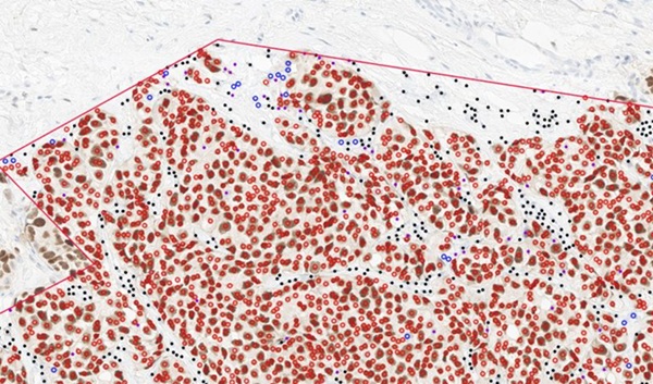
Researchers at Tulane University (New Orleans, LA, USA) are leading a project called MAGIC-SCAN (Machine-learning Assisted Gigantic Image Cancer margin SCANner) which aims to become one of the fastest high-resolution tissue scanners in the world. This system would be capable of identifying residual cancer cells on the surface of excised organs in a matter of minutes. The scanner would be trained on a vast database of clinical scans to accurately identify cancer cells at a cellular level, producing a detailed 3D map of the tumor’s surface. The new technology combines advances in microscopy, automation, computing, and machine learning, utilizing optical-sectioning super-resolution structured illumination microscopy to achieve twice the resolution of conventional microscopes.
.jpg "Project leads J. Quincy Brown (left), associate professor of biomedical engineering, and Brian Summa, associate professor of computer science, test a prototype of a new imaging system (Photo courtesy of Tulane University)")
This cutting-edge imaging tool promises to transform cancer surgery by enabling doctors, while the patient is still under anesthesia, to verify the complete removal of cancer, potentially eliminating the need for additional surgeries. The Tulane research team has been developing this technology with a focus on prostate and colorectal cancers—two of the most challenging tumors to excise—reducing detection time to approximately 45 minutes. They have built a prototype of this groundbreaking system and are now leading efforts to address the remaining technical, computing, and engineering challenges to actualize this device within five years. Efforts are underway to enhance imaging resolution quality and develop the necessary cyberinfrastructure to manage extensive data sets required for training the machine-learning models. Furthermore, the team plans to conduct clinical validation of the device and develop versions compliant with FDA regulations.
“Currently, it can take days to weeks before a surgeon knows whether all the tumor has been removed, and our goal is to get that down to 10 minutes, while the patient is still on the table,” said J. Quincy Brown, PhD, associate professor of biomedical engineering in the Tulane School of Science and Engineering and lead researcher on the project. “If successful, our work would transform cancer surgery as we know it.”
Related Links:
Tulane University