New Method Aligns Data from Tissue Slices Virtually to Expand Possibilities for 3D Analysis
Posted on 22 Aug 2023
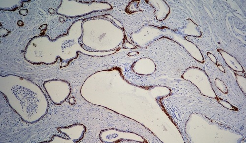
When it comes to studying biological tissue, whether from a patient or an animal, a common approach involves surgically removing a portion of the affected tissue for analysis. In laboratories across the globe, technicians meticulously slice the extracted tissue into thin sections, which are then examined under a microscope or subjected to tests that detect specific molecules. These molecular clues can aid in diagnosing conditions, guiding treatments, or even gauging the effectiveness of drugs. However, the process of analyzing each slice is resource-intensive in terms of time, money, and computational capacity. Consequently, researchers and medical professionals are often constrained to studying only a limited number of slices from various parts of the tissue. Adding to the complexity, the act of cutting, processing, and analyzing tissue slices within a lab environment causes them to undergo physical distortions. As a result, accurately understanding how these slices align and fit into the three-dimensional structure of the original tissue becomes challenging. Now, a new method revolutionizes the ability to comprehend the three-dimensional composition of tissues, including tumors or other tissue using data from just a few slices, thus allowing for a much deeper understanding of biological tissue samples.
The new method, named Gaussian Process Spatial Alignment (GPSA) which has been developed by researchers at Gladstone Institutes (San Francisco, CA, USA) has a scope extending beyond tumors, encompassing a wide array of tissues and data derived from tissue slices. This innovative method leverages a two-layer Gaussian process. In the first layer, it aligns the warped two-dimensional tissue slice onto a three-dimensional tissue model. The second layer attributes data collected from the slice, such as activated genes, to each point in the three-dimensional model. This intelligent approach effectively reverses the warping effect, resulting in a precise alignment of the slices. During this process, the GPSA model extrapolates data to fill the gaps between slices, generating a comprehensive three-dimensional "atlas" of the tissue.
")
A key advantage of GPSA is its versatility. Researchers can construct tissue atlases using data from slices of varying sizes, generated by diverse technologies, and captured at different scales and resolutions. Unlike previous techniques that demanded a predefined three-dimensional framework, GPSA derives this framework solely from the two-dimensional slices when an initial framework isn't available. Additionally, GPSA has the capability to merge multiple types of tissue-slice data, such as genetic activity and cellular structure, into a unified atlas. Furthermore, when applied to slices taken from the same tissue at different time points, GPSA can generate atlases that predict how each location within the tissue evolves over time. This feature could deepen our insights into aging, disease progression, and the development of different tissues within growing organisms. Presently, researchers are engaged in further analyses to further demonstrate the tool’s flexibility. For instance, they have devised an approach that budget-conscious labs can use to determine the minimum number of tissue slices required and the precise cutting locations needed for GPSA to build an informative tissue atlas.
“Say you have four slices from different locations in a person’s breast cancer tumor, and for every point on each slice you know which of 20,000 genes are turned on or off,” said Gladstone Senior Investigator Barbara Engelhardt, PhD, senior author of the study. “GPSA creates a fully query-able 3D atlas where, for any single ‘x, y, z’ coordinate, for any of the 20,000 genes, we can dive in and ask: What genes are on and off at this position in the tumor? And how certain are we in this estimate?”
Related Links:
Gladstone Institutes
 Assay.jpg)