AI Tool Helps Make Real-Time Diagnosis During Surgery
Posted on 26 Dec 2022
When a patient undergoes a surgical operation to remove a tumor or treat a disease, the course of surgery is often not predetermined. To decide how much tissue needs to be removed, surgeons must know more about the condition they are treating, including a tumor’s margins, its stage and whether a lesion is malignant or benign - determinations that often hinge upon collecting, analyzing, and diagnosing a disease while the patient is on the operating table. When surgeons send samples to a pathologist for examination, both speed and accuracy are of the essence. The current gold-standard approach for examining tissues often takes too long and a faster approach, which involves freezing tissue, can introduce artifacts that can complicate diagnostics. Now, researchers have developed a new method that leverages artificial intelligence to translate between frozen sections and the gold-standard approach, thereby improving the quality of images to increase the accuracy of rapid diagnostics.
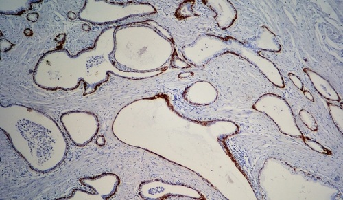
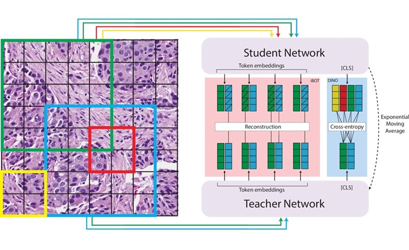
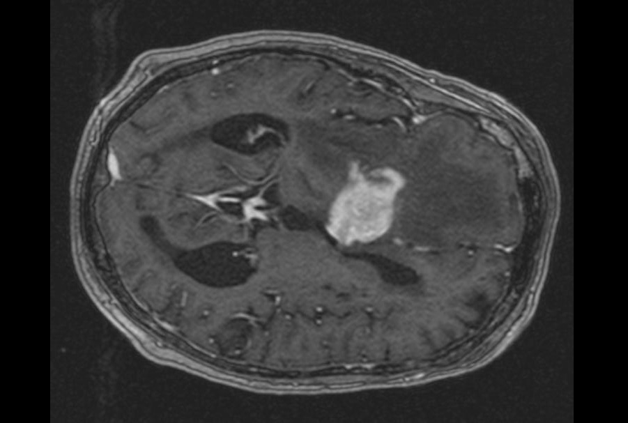
For making final diagnoses, pathologists use formalin-fixed and paraffin-embedded (FFPE) tissue samples - this method preserves tissue in a way that produces high-quality images but the process is laborious and typically takes 12 to 48 hours. For a rapid diagnosis, pathologists use an approach known as cryosectioning that involves fast freezing tissue, cutting sections, and observing these thin slices under a microscope. Cryosectioning takes minutes rather than hours but can distort cellular details and compromise or tear delicate tissue. Researchers at the Brigham and Women’s Hospital (Boston, MA, USA) have developed a deep-learning model that can be used to translate between frozen sections and more commonly used FFPE tissue. The team demonstrated that the method could be used to subtype different kinds of cancer, including glioma and non-small-cell lung cancer.
")
The researchers validated their findings by recruiting pathologists to a reader study in which they were asked to make a diagnosis from images that had gone through the AI method and traditional cryosectioning images. The AI method not only improved image quality but also improved diagnostic accuracy among experts. The algorithm was also tested on independently collected data from Turkey. The researchers note that in the future, prospective clinical studies should be conducted to validate the AI method and determine if it can contribute to diagnostic accuracy and surgical decision-making in real hospital settings.
“We are using the power of artificial intelligence to address an age-old problem at the intersection of surgery and pathology,” said corresponding author Faisal Mahmood, PhD, of the Division of Computational Pathology at BWH. “Making a rapid diagnosis from frozen tissue samples is challenging and requires specialized training, but this kind of diagnosis is a critical step in caring for patients during surgery.”
“Our work shows that AI has the potential to make a time-sensitive, critical diagnosis easier and more accessible to pathologists,” said Mahmood. “And it could potentially be applied to any type of cancer surgery. It opens up many possibilities for improving diagnosis and patient care.”
Related Links:
Brigham and Women’s Hospital