Breast Cancer Histopathology Employs Infrared Spectroscopic Imaging
By LabMedica International staff writers
Posted on 19 Apr 2021
Digital analysis of cancer specimens using spectroscopic imaging coupled to machine learning is an emerging area that links spatially localized spectral signatures to tissue structure and disease. Breast histopathology, as an example of the broad relevance of these techniques, is critically important for clinical diagnoses. Posted on 19 Apr 2021
Current histologic characterization is morphology-based; thin tissue sections are stained, and cells are visually recognized by a pathologist using an optical microscope. However, the basis of the disease is well known to be molecular. Molecular analysis for pathology is complicated by the spatial diversity of cells and acellular materials, necessitating an analytical technique that involves imaging.
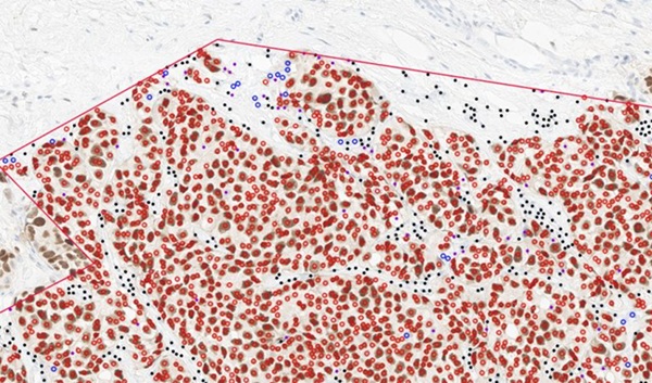
 and standard definition (SD) classification performance using the 6-class model (Photo courtesy of University of Illinois at Urbana−Champaign)")
Image: Spatial and quantitative comparison of high-definition (HD) and standard definition (SD) classification performance using the 6-class model (Photo courtesy of University of Illinois at Urbana−Champaign)
Bioengineers at the University of Illinois at Urbana−Champaign (Urbana, IL, USA) and their colleagues examined the role of spatial-spectral tradeoffs in infrared spectroscopic imaging configurations for probing tumors and the associated microenvironment profiles at different levels of model complexity. The imaged breast tissue using standard and high-definition Fourier Transform Infrared (FT-IR) imaging and systematically examine the localization, spectral origins, and utility of data for classification.
The team obtained formalin-fixed, paraffin-embedded serial breast tissue microarrays (TMA) sections. The array consisted of a total of 101 cores of 1 mm diameter from 47 patients. Two sections were stained with hematoxylin and eosin (H&E) and other immunohistochemical markers and imaged with a light microscope. High-definition (HD) FT-IR imaging was conducted using the Agilent Stingray imaging system (Santa Clara, CA, USA) which is comprised of a 680-IR spectrometer coupled to a 620-IR imaging microscope with 0.62 numerical aperture, 25×objective.
The scientists provided a systematic comparison in the use of HD and SD FT-IR imaging data for breast pathology in their study. While the increased spatial localization of spectral signals in HD imaging may have been expected to provide a confounding influence, the study demonstrated that accuracy can be high, and there is significant potential in this sampling mode offering higher sensitivity. The team stated that IR imaging can not only provide the recognition capability of molecular data but can also balance that with an increased quality of morphologic data.
Rohit Bhargava, PhD, bioengineering professor and senior author of the study, said, “As technology expands and provides more capabilities with new features, it becomes more difficult to choose the optimal technology from the many options available. This study provides a nice comparison and guidelines to design a more useful and practical technology.” The study was originally published on February 27, 2021 in the journal Clinical Spectroscopy.
Related Links:
University of Illinois at Urbana−Champaign
.jpg)
Gold Member
Clinical Chemistry Assay
Sorbitol Dehydrogenase (SDH)


Food Allergy Screening ELISA Kit
Allerquant 14G B ELISA

Creatinine/eGFR Meter
StatSensor® Creatinine/eGFR Meter