Roche Advances Personalized Healthcare with Development of Image Analysis Algorithms Using Artificial Intelligence
By LabMedica International staff writers
Posted on 29 Jun 2020
Roche (Basel, Switzerland) has announced the CE-IVD launch of its automated digital pathology algorithm, the uPath PD-L1 (SP263) image analysis for non-small cell lung cancer (NSCLC). Posted on 29 Jun 2020
Roche is delivering the end-to-end digital pathology solution from tissue staining to producing high-quality digital images that can be reliably assessed using automated clinical image analysis algorithms. Roche’s uPath image analysis algorithm suite for pathology decision support offers ready-to-use image analysis tools, providing fast, consistent and automated analysis so that pathologists can quickly, accurately and confidently assess immunohistochemistry/in situ hybridization and hematoxylin and eosin-stained slides. All algorithms in the suite for uPath software will provide image analysis of VENTANA DP 200 scanned slide images stained with a Roche tissue assay. Together, Roche is delivering a new foundation of its digital pathology solution which will enable the development of artificial intelligence-based image analysis algorithms that can provide pathologists more tools to improve efficiency and precision.
 image analysis (Photo courtesy of Roche)")
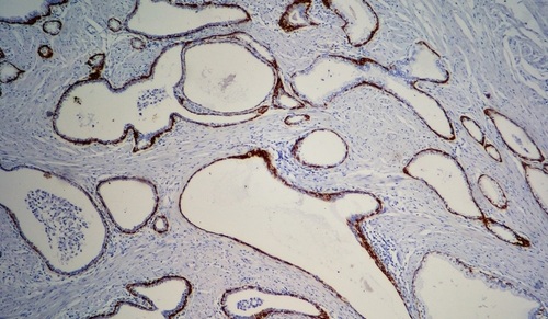
Image: uPath PD-L1 (SP263) image analysis (Photo courtesy of Roche)
The uPath PD-L1 (SP263) image analysis for NSCLC algorithm’s whole-slide automated analysis uses artificial intelligence to provide, with one-click, assessments of scanned slide images that are objective and reproducible and have the potential to aid diagnosis and, ultimately, targeted treatment options for patients. Validated on the VENTANA PD-L1 (SP263) Assay, the algorithm is ready-to-use and integrated within the Roche uPath enterprise software, a universal digital platform for case management, collaboration and reporting. The algorithm will help pathologists to quickly determine whether tumors are positive for the PD-L1 biomarker, highlighting positively and negatively stained tumor cells with a clear visual overlay for easy reference. It is intended for in vitro diagnostic use as an aid to the pathologist in the display, detection, counting, review and classification of tissues and cells of clinical interest based on particular morphology, color, intensity, size, pattern and shape. Patients with tumors that are positive for the PD-L1 biomarker may be eligible for targeted treatment.
“Improving diagnostic consistency and certainty is crucial in providing faster, higher-quality and more accurate diagnoses to cancer patients,” said Thomas Schinecker, CEO, Roche Diagnostics. “Our uPath PD-L1 (SP263) image analysis for non-small cell lung cancer is the first next-generation CE-IVD PD-L1 algorithm to the clinical market. It expands on our growing digital pathology suite for VENTANA assays that aid physicians in providing the most accurate treatment decisions for patients with the most common type of lung cancer.”
 Assay.jpg)
Gold Member
H-FABP Assay
Heart-Type Fatty Acid-Binding Protein Assay


Automated Clinical Chemistry Analyzer
Envoy 500+

New
Gold Member
Pre- Eclampsia Control
Acusera Pre-Eclampsia Control