Diagnostic Biomarker Found for Esophageal Squamous Cell Carcinoma
By LabMedica International staff writers
Posted on 22 Jan 2020
Esophageal cancer is one of the most common malignant tumors in the world, and its incidence ranks seventh among those of all malignant tumors. Esophageal cancer can be divided into two pathological types: squamous cell carcinoma (SCC) and adenocarcinoma.Posted on 22 Jan 2020
Leucine‐rich repeat‐containing G protein–coupled receptor (LGR) plays a pivotal role in adult stem cells, which are markers of various types of adult stem cells in the skin, nails, and a group of basal and intraluminal progenitors that induce luminal tumorigenesis. LGR6 can promote the self‐renewal and progression of non–small‐cell lung cancer and has strong carcinogenic potential.
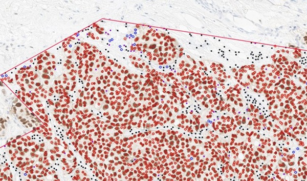
 in esophageal squamous cell carcinoma (ESCC) and normal esophageal tissues (NT) (Photo courtesy of Fujian Medical University).")
Image: Histological immunostaining showing expression of leucine‐rich repeat‐containing G protein–coupled receptor 6 (LGR6) in esophageal squamous cell carcinoma (ESCC) and normal esophageal tissues (NT) (Photo courtesy of Fujian Medical University).
Scientists at the Fujian Medical University Union Hospital (Fuzhou, China) collected during surgical resection of esophageal cancer, 102 Esophageal Squamous Cell Carcinoma (ESCC) samples and their corresponding non‐tumor esophageal tissues. Tissues were immediately frozen in liquid nitrogen and stored in a −80 °C freezer or fixed in 10% formalin for paraffin embedding.
Total RNA was extracted from frozen tissue and 1 mg of RNA was reverse transcribed for first complementary DNA strand synthesis using a miScript Reverse Transcription Kit (Qiagen, Hilden Germany). Real‐time quantitative polymerase chain reaction (PCR) was performed using a SYBR Premix EX Taq Kit (TakaraBio, Shiga, Japan). The relative mRNA expression of LGR6 was detected with the 2(-Delta Delta C(T)) method using specific primers and its expression level was normalized to that of endogenous β‐actin. Other techniques used by the investigators included Western Blots Analysis and immunohistochemistry (IHC).
The scientists reported that the expression of LGR6 in ESCC tissues was significantly higher than that in normal tissues and was negatively correlated with the differentiation degree of ESCC and the prognosis of the patients but not closely correlated with the Classification of Malignant Tumors (TNM) stage of ESCC. Protein‐protein interaction (PPI) networks showed that LGR6 had a close interaction with R-spondin-1 (RSPO1), RSPO2, RSPO3, and RSPO4. Kyoto encyclopedia of genes and genomes (KEGG) pathway analysis showed that LGR6 activated the Wnt/β‐catenin signaling pathway by binding with RSPO ligands to promote the progression of ESCC.
The authors concluded that the study confirmed for the first time that LGR6 is highly expressed in ESCC tissues and that increased expression of LGR6 is associated with a poor prognosis of ESCC patients. These findings provide a basis for the potential application of LGR6 as a biomarker for early diagnosis and as a target gene for early therapeutic intervention. The study was published on January 9, 2020 in the Journal of Clinical Laboratory Analysis.
Related Links:
Fujian Medical University Union Hospital
Qiagen
TakaraBio

New
Gold Member
Pre- Eclampsia Control
Acusera Pre-Eclampsia Control


Thyroid Test
Anti-Thyroid EIA Test

Electrolyte Analyzer
BKE-B