Cancer Cells Detected Before They Form Metastases
By LabMedica International staff writers
Posted on 06 Dec 2015
A versatile microarray-based platform has been developed that is able to capture single target cells from large background populations as analyses of rare events occurring at extremely low frequencies in body fluids are still challenging.Posted on 06 Dec 2015
Single cancer cells migrate with blood flow through the body before they settle in new tissue and in this way, metastases may be formed, even after the main tumor was treated successfully. It is difficult to detect cancer cells in the blood at an early stage as about one malignant cell is encountered per billion of healthy cells.

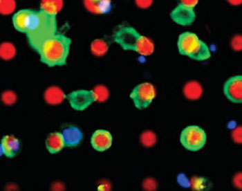
Image: A micrograph of the target cells (green) sticking to the microarray platform (red) (Photo courtesy of Dr. Michael Hirtz / KIT).
Scientists at the Karlsruhe Institute of Technology (Germany) and their colleagues fabricated the device by means of polymer pen lithography, a surface is provided with a microscopically small structure using a plastic die. The target cells adhere to these structures. The blood sample to be investigated is injected into a flat microchannel that crosses the platform. As a maximum number of target cells are to contact the array, a fishbone-shaped structure at the top of the channel stirs up the passing liquid.
In order to underline the feasibility of the presented system in the clinical settings, a series of blood samples from nine breast and one colon cancer patient was analyzed. Samples from healthy individuals were used as negative controls. Prior to pumping the blood sample through the microfluidic device, circulating tumor cells (CTC’s) were pre-enriched by size filtration using the Parsortix System (ANGLE plc; Guilford, UK) using 4 mL of blood per patient mainly to extract red blood cells and to reduce the number of leukocytes. Cells larger than 10 μm in diameter were captured by the Parsortix system, harvested, and exposed to an antibody cocktail of biotinylated anti-EpCAM and anti-HER2 antibodies.
CTCs were discovered in six out of 10 blood samples using the micropattern platform with a range from 0.75 to 2 CTCs/mL. Four samples were deemed negative since both CellSearch (Janssen Diagnostics, LLC; South Raritan, NJ, USA) and the microarray approach were not able to find any CTCs. In three of the remaining six blood samples CellSearch found more CTCs than captured by the new system, range from 3.7to 7.6 CTCs/mL by CellSearch compared to 1.25 to 2 CTCs/mL by the micropattern platform. Two blood samples from patients were even tested positive by the novel device, whereas CellSearch was negative.
Michael Hirtz, ScD, a professor and senior author of the study said, “With our method, we reach a very high hit rate: more than 85% of the extracted cells really are cancer cells, In addition, we can sample suspicious cells undamaged and study them in more detail. While the tumor cells dock to the prepared locations according to the key-lock principle, the remaining cells are simply washed away.” The study was published on October 23, 2015, in the journal Scientific Reports.
Related Links:
Karlsruhe Institute of Technology
ANGLE plc
Janssen Diagnostics


