Breast Cancer Diagnosis Uses Spatial Light Interference Microscopy
By LabMedica International staff writers
Posted on 17 Sep 2015
The standard practice in histopathology of breast cancers is to examine a hematoxylin and eosin (H&E) stained tissue biopsy under a microscope to diagnose whether a lesion is benign or malignant.Posted on 17 Sep 2015
A new optical method called Spatial Light Interference Microscopy (SLIM) has been used to quickly and accurately determine whether breast tissue lesions are cancerous, as this quantitative, label-free, and high-throughput diagnosis method is highly advantageous.
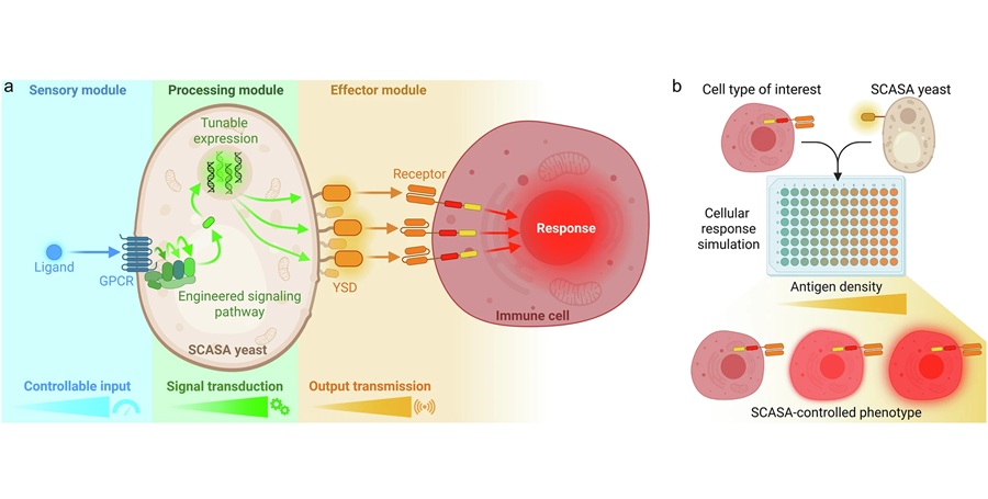
 and SLIM (bottom row) images in their respective abilities to show malignant and benign. The images were obtained from adjacent sections. Color bars are in radians (Photo courtesy of Hassaan Majeed).")
Image: A comparison of stained bright-field microscopy (top row) and SLIM (bottom row) images in their respective abilities to show malignant and benign. The images were obtained from adjacent sections. Color bars are in radians (Photo courtesy of Hassaan Majeed).
Scientists at the University of Illinois (Urbana, IL, USA) used the breast tissue biopsies of 400 different patients, and selected two parallel, adjacent sections from each biopsy. One was stained and the other left unstained. The unstained samples were analyzed using a SLIM module attached to a commercial phase contrast microscope to generate interferograms, which are photographic images derived from data based on how the tissue refracts light.
Four interferograms were used to produce one quantitative image showing areas with different refractive properties in different colors. The boundary between tumors and the cells around them were clearly delineated, making it possible to assess whether the tumors were malignant or benign. As a first step toward quantitative diagnosis based on SLIM, the team carried out a qualitative evaluation of our label-free images. These images were shown to two pathologists who classified each case as either benign or malignant. This diagnosis was then compared against the diagnosis of the two pathologists on corresponding H&E stained tissue images and the number of agreements were counted.
The agreement between SLIM and H&E based diagnosis was 88% for the first pathologist and 87% for the second. The results demonstrate the potential and promise of SLIM for quantitative, label-free, and high-throughput diagnosis. YongKeun Park, PhD, a professor at Korean Advanced Institute of Science and Technology (Daejeon, Republic of Korea) and a guest editor of the of the special section on Quantitative Phase Imaging in Biomedicine in which the study appears said, “Conventional methods for diagnosis of breast cancer have several limitations, including observer discrepancy.” The study was published on August 20, 2015, in the Journal of Biomedical Optics.
Related Links:
University of Illinois
Korean Advanced Institute of Science and Technology

Gold Member
Respiratory Syncytial Virus Test
OSOM® RSV Test


Hemodynamic System Monitor
OptoMonitor
Pipette
Accumax Smart Series