New Diagnostic Tool on Horizon for Surgeons Treating Cancer Patients
By LabMedica International staff writers
Posted on 06 Jul 2015
Researchers have successfully developed a new tool that may enable surgeons to determine if a biopsy tissue is cancerous while their patients are still on the operating table, without routinely requiring microscope-based pathology analysis of the tissue. Posted on 06 Jul 2015
The tool, developed by a team of researchers from the Department of Energy (DOE)'s Oak Ridge National Laboratory (ORNL; Oak Ridge, TN, USA) and the Harvard Medical School teaching hospital Brigham & Women’s Hospital (Boston, MA, USA), is a new mass spectrometry (MS)-based technology—an automated droplet-based surface-sampling probe that accomplishes in about 10 minutes what currently takes 20–30 minutes. First author Dr. Vilmos Kertesz of ORNL expects the time to soon be further cut to 5 minutes.
.")
Image: The droplet-based surface sampling probe, a newly developed mass spectrometry-based technology, speeds the process of analyzing a cancer patient liver biopsy sample (Photo courtesy of Oak Ridge National Laboratory).
.")
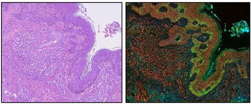
Image: A droplet-based surface sampling probe mass spectrometry diagnostic tool was successfully used in a proof-of-concept study profiling of hormones in human pituitary gland and tumor thin tissue sections (Photo courtesy of Kertesz V et al., 2015, and the journal Analytical and Bioanalytical Chemistry).
The system has been successfully used for spatially resolved sampling and detection of drugs and metabolites from thin sections of animal tissue as well as of proteins from dried blood. For the proof-of-concept study, the researchers rapidly profiled two hormones from human pituitary tissue.
“Instead of having to cut and mount tissue and wait for a trained pathologist to review the sample under a microscope, a technician might soon perform an equally conclusive test in the operating environment,” said Dr. Kertesz. The technology may also become an attractive alternative to the traditional diagnostic method of biomarker testing with immunohistochemistry (IHC). Although IHC provides a high degree of spatial recognition, it is time consuming and is limited by the quality and specificity of the antibody.
The success of this work can be traced back to patents resulting from previous DOE projects and it advances the liquid microjunction surface sampling probe technology first patented by ORNL. Currently ORNL houses the only laboratories worldwide that have this automated droplet-based surface sampling probe and the requisite software.
While other MS-based techniques (such as desorption electrospray ionization and rapid evaporative ionization) are being evaluated for classifying tumors and providing prognostic information, they are limited mainly to analysis of lower molecular weight biomolecules. The new droplet-based method overcomes this limitation. “The ability to quickly characterize the tissue distribution of larger macromolecular biomarkers like peptides and proteins would harness the diagnostic value of validated IHC approaches for surgical decision-making,” said Dr. Kertesz, “On the basis of the results and the relative simplicity, rapidity, and specificity of our method, there is great potential for our technology to assist surgeons in the detection of cancer from tissue biopsy samples.”
The study, Kertesz V et al., was published June 18, 2015, in the journal Analytical and Bioanalytical Chemistry.
Related Links:
Oak Ridge National Laboratory
Brigham & Women’s Hospital

Platinum Member
COVID-19 Rapid Test
OSOM COVID-19 Antigen Rapid Test


Anti-Cyclic Citrullinated Peptide Test
GPP-100 Anti-CCP Kit

New
Gold Member
TORCH Panel Rapid Test
Rapid TORCH Panel Test