Early-Stage Cancers May Be Detected with Noninvasive Prenatal Testing
By LabMedica International staff writers
Posted on 24 Jun 2015
A noninvasive prenatal testing (NIPT) is a screening technique used to detect Down syndrome and other conditions involving chromosomal abnormalities in a developing fetus. Posted on 24 Jun 2015
Similar to placental DNA, tumor DNA can be detected in the plasma, and analysis of cell-free tumor DNA can be used to characterize and monitor cancers, and profiling plasma DNA allows for presymptomatic detection of tumors in pregnant women undergoing routine NIPT.
.")
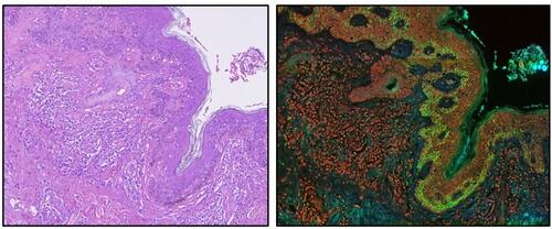
Image: Histopathology of classical Hodgkin lymphoma (Photo courtesy of Dr. John K.C. Chan).
Gynecological oncologists at the Katholieke Universiteit (KU) Leuven–University of Leuven (Belgium) optimized a large parallel sequencing–based NIPT dataset and analysis, which not only interrogates the common trisomies but also allows the genome wide discrimination of fetal and maternal segmental aneuploidies. The investigators analyzed more than 6,000 pregnant women using an adapted version of NIPT, and the team identified three genomic abnormalities in three of the women that they could not link to maternal or fetal profiles.
To confirm that the abnormal genomic representation (GR) profile was due to tumor-derived cell-free DNA (cfDNA), fluorescence in situ hybridization (FISH) was performed on tumor biopsy using probes for different genes which confirmed that the genomic imbalances identified in the cfDNA matched the gains and losses of the corresponding chromosomal regions in carcinoma cells The analysis uncovered the presence of an ovarian carcinoma, a follicular lymphoma, and a Hodgkin lymphoma.
The woman in whom follicular lymphoma was identified was found to have inactive cancer, so no treatment was required. Chemotherapy was given to the other two women, however, with one being treated during pregnancy. She subsequently gave birth to a healthy baby girl. Follow-up assessments in the women who underwent chemotherapy allowed the oncologists to monitor the effectiveness of treatment, which showed that during and after chemotherapy, the genomic profiles of the women returned to normal.
Nathalie Brison, PhD, a coauthor of the study said, “We now know that it is possible to offer the accurate detection of chromosomally imbalanced cancers to the general population via minimally invasive screening methods. The normalization of the NIPT profile in these patients following treatment indicates that we can also measure response to treatment as early as after the first administration of chemotherapy.” The study was published on June 5, 2015, in JAMA Oncology.
Related Links:
Katholieke Universiteit (KU) Leuven–University of Leuven

Platinum Member
COVID-19 Rapid Test
OSOM COVID-19 Antigen Rapid Test


Anti-Cyclic Citrullinated Peptide Test
GPP-100 Anti-CCP Kit

Gold Member
Real-time PCR System
GentierX3 Series