Gene Expression Correlates with Lymphocyte Infiltration in Breast Cancer
By LabMedica International staff writers
Posted on 31 Dec 2014
Lymphocytic infiltration is associated with a favorable prognosis and predicts response to chemotherapy in many cancer types, including the aggressive triple-negative breast cancer (TNBC).Posted on 31 Dec 2014
A novel high-tech system has been developed for measuring the body's immune response to cancer as a way of assessing how rapidly the disease is likely to progress. The system combines computerized imaging of tumor samples with statistical analysis, and is the first objective method to measure the interaction between a patient's immune system and their tumor.

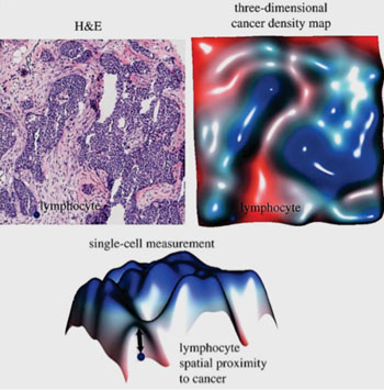
Image: A histology slide stained with hematoxylin and eosin (H&E) and the corresponding three-dimensional cancer density map, which facilitate the measurement of spatial proximity to cancer for every single lymphocyte in the image (Photo courtesy of The Institute of Cancer Research).
Scientist at The Institute of Cancer Research (London, UK) analyzed 181 samples from women with triple-negative breast cancer. On average, three tumor sections were obtained from different locations of each primary tumor and placed onto the same slide. Tumor materials sandwiched between these sections were sectioned, mixed and used for molecular profiling, thereby maximizing the biological relevance of multiple data types being generated.
The team developed a system, which split lymphocytes into three classes depending on their location within the tumor, and calculated how many of each type were present in each sample, and how many cancer cells were present. Immune infiltration was scored for 112 of the 181 samples by the pathologists into three categories: absent, mild and severe: absent if there were no lymphocytes, mild if there was a light scattering of lymphocytes and severe if there was a prominent lymphocytic infiltrate. Gene expression data were profiled using the human linkage-12 genotyping beadchip (HT-12) platform (Illumina; San Diego, CA, USA).
Women with fewer than around eleven intra-tumor lymphocytes per 1,000 cancer cells had an average five-year survival rate of 49%, compared with an average of 80% in triple-negative breast cancer as a whole. They also found a correlation between immune infiltration of tumors and increased levels of a protein called cytotoxic T-lymphocyte-associated protein 4, (CTLA4) or cluster of differentiation 152 (CD152), suggesting it could be a potential treatment target in this breast cancer type.
Yinyin Yuan, PhD, the team leader of the study, said, “Our test combines imaging technology with computerized analysis of large amounts of data from tumor samples, which typically contain more than 100,000 cells. We found the technique could accurately identify high-risk tumors that were evading the body's immune system in this type of breast cancer, and hope it can be adapted and added to doctors' arsenal against a variety of cancers.” The study was published on December 10, 2014, in the Journal of The Royal Society Interface.
Related Links:
The Institute of Cancer Research
Illumina


.jpg)
