Digital Slide Imaging Evaluated for Cervicovaginal Cytology
By LabMedica International staff writers
Posted on 26 Jun 2013
Digital imaging is the creation, storage, and transmission of an image file using a computer and is considered by many to be the future of anatomic pathology.Posted on 26 Jun 2013
Whole slide imaging has been successfully used in surgical pathology, but its usefulness and clinical application have been limited in cytology for several reasons, including lack of availability of z-axis depth focusing and large file size.
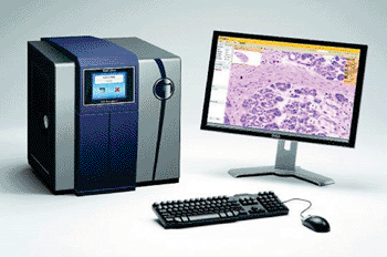
Image: BioImagene digital scanner (Photo courtesy of Ventana Medical Systems).
Cytopathologists at The Methodist Hospital (Houston, TX, USA) investigated the accuracy and efficiency of whole slide imaging, as compared with traditional glass slides, for use in cervicovaginal diagnostic cytology. Eleven cervicovaginal cytology cases were evaluated by four cytotechnologists and three cytopathologists in a blinded study. Digitally scanned images were reviewed first, and different magnification scans were presented as separate cases in a random order. Corresponding glass slides were also reviewed and for each case, the diagnoses and total time to reach each diagnosis were recorded.
The specimens were prepared using both ThinPrep (BD Diagnostics TriPath; Burlington, NC, USA) and SurePath (Hologic Inc.; Marlborough, MA, USA). All the cases were scanned using the BioImagene iScan Coreo Au 3.0 scanner (Ventana Medical Systems Inc.; Sunnyvale, CA, USA). Diagnostic accuracy was higher and average time per case was lower with glass slides as compared with all digital images. Accuracy of interpretation of the digital images was best achieved with ×40 magnification, and z-stack digital images scanned at ×40 magnification improved the accuracy of diagnosis in one case of adenocarcinoma of the endometrium.
The authors concluded that whole slide imaging is a viable option for the purposes of teaching and consultations, and as a means of archiving cases. However, considering the large file size and total time to reach diagnosis on digital images, they consider whole slide imaging may not yet be ready as a daily screening tool in cervicovaginal diagnostic cytology. The study was published in the May 2013 edition of the journal Archives of Pathology & Laboratory Medicine.
Related Links:
The Methodist Hospital
BD Diagnostics
Ventana Medical Systems




Platinum Member

Automated Coagulation Analyzer
Hemolumi H6

Gold Member

Aspiration System
VACUSAFE
New

Fully Automated Urinalysis System
DxU 1800 Fully Automated Urinalysis System
New

Automated Immunoassay Analyzer
SuperFlex™