Breast Cancer Cells Characterized by Microspectroscopy
By LabMedica International staff writers
Posted on 05 Nov 2012
A diagnostic tool has been developed that identifies the metastatic ability of breast cancer cells based on the characterization of the lipid component of the cells. Posted on 05 Nov 2012
The characterization of the lipids associated with malignancy has been possible due to technological development of a spectroscopic device named Raman (RS) and forms the basis for introducing this technique in routine cytological diagnosis.
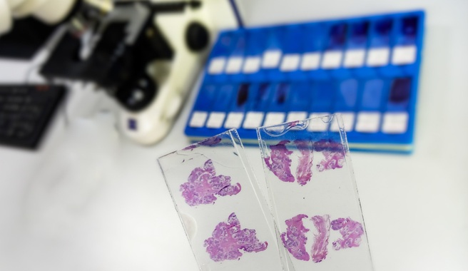
Scientists at the Bellvitge Biomedical Research Institute (IDIBELL; Barcelona, Spain) assessed the different profiling of the lipid composition of breast cancer cells, which permitted differentiation of the lipogenic phenotype according to the proportion of unsaturated fatty acids. The investigators cultured breast cancer cells and applied immunochemistry, before submitting the samples to Raman microspectroscopy
The Renishaw Raman system (Apply Innovation; Wotton-under-Edge, UK) comprises of a 514 nm laser that supplies an excitation beam of about 10 mW power, which is focused onto the sample via a microscope. The cytoplasm lipids were measured by RS in a position near the nucleus and outside the endoplasmic reticulum area, where the Nile red staining showed major lipid concentration. The lipid phenotype associated to breast cancer malignancy belongs to Raman spectra acquired in the range of 2,820-3,030 cm−1.
The authors concluded that Raman spectroscopy is a promising technique in biomedical studies due to its noninvasive character and high specificity. The cytology techniques used have found a correlation between the activation of lipogenesis, the chemical reaction leading to fatty acids in an organism, and the amount of saturated fats in metastatic cells indicating a worse prognosis and a decreased survival. The lipid content of the breast cancer cells might be a useful measure to determine various functions coupled to the progression of breast cancer. Àngels Sierra, PhD, the senior author, said, “The algorithm for the discrimination of the metastatic ability is a first step towards the stratification of breast cancer cells using this quick and reactive tool." The study was published on October 17, 2012, in the journal Public Library of Science ONE.
Related Links:
Bellvitge Biomedical Research Institute
Apply Innovation

Gold Member
Automatic Hematology Analyzer
CF9600


Electrolyte Analyzer
CBS-4000 (CBS-400)

Pipette Calibration System
Artel PCS®