“Virtual Slides” Reveal Disease Tissue in 3D
By LabMedica International staff writers
Posted on 07 May 2012
A fast, user-friendly system has been developed for examining tissue samples in “virtual” 3D images. The novel digital scanning 3D reconstruction system produces high-resolution, multicolored images that can be rotated and examined from any angle. Posted on 07 May 2012
Computing experts and medical researchers at the University of Leeds (Leeds, UK) have combined efforts to developed this technology as a particularly useful tool for histopathology researchers and potentially also for clinical practice, as medical imaging technology provides even higher resolution images of tissue.
.")
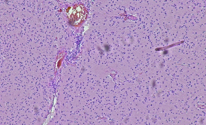
Image: Studying tissue samples in “virtual” 3D -- Please see Related Links below for additional images (Photo courtesy of the University of Leeds).
Viewing tissue in 3D enables more in-depth understanding of tissue shape characteristics in ways not possible with conventional methods. Currently, hospital pathologists and medical researchers cut tissue samples into ultra-thin slices and routinely examine these by hand, one-by-one, on a microscope. This is a labor-intensive process - a single slide can contain several hundred thousand cells. To perform a 3D-like analysis, users would need to look at hundreds of different 2D sections - something that would be prohibitively expensive and time-consuming.
In contrast, the system developed at the University of Leeds requires almost no extra manual input once the tissue has been cut and mounted onto glass slides. An automated system turns batches of the slides into high-resolution digital images, which are then aligned using image registration software. Users, without input from computing specialists, can then study these virtual blocks of tissue in 3D and zoom in on particular areas of interest.
The researchers have now tested the system on eight different types of tissue, using more than 13,000 virtual slides to create around 400 separate 3D volumes. The system and selected case studies, including examples of liver disease, cancer, and embryology, are described in the May 2012 issue of the American Journal of Pathology.
This new approach to digital 3D reconstruction reveals more detailed information about disease processes - information that could be used to develop new therapies or explain why conventional treatments are not working. "Having a 3D view can often make a real difference," said Dr. Derek Magee, from the University of Leeds School of Computing, where the system’s software was developed. "For instance, if you want to understand how a system of blood vessels supplying a tumor connects up, you really need to see that in 3D, not as a series of separate 2D sections."
Related Links:
University of Leeds
Sample 3D images

Gold Member
STI Test
Vivalytic MG, MH, UP/UU


Hematology Consumables
Bioblood Devices

Prefilled Tubes
Prefilled 5.0ml Tubes