


Organ-on-Chip System Offers Insights Into Tumor Interactions and Treatment Response
Posted on 16 Nov 2026
Solid tumors interact closely with blood vessels, nerves, and surrounding tissues, making it difficult to study tumor growth, invasion, pain, and treatment response under controlled laboratory conditions. Organ-on-chip technology offers a way to recreate key aspects of these complex interactions outside the body. Using this approach, researchers have developed a human organ-on-chip model to investigate how cancer interacts with its surrounding microenvironment.
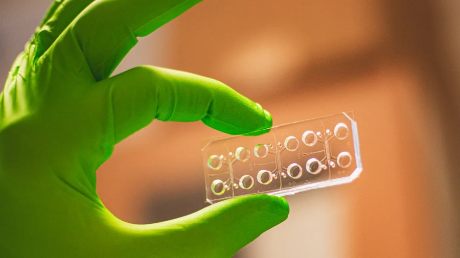
Researchers at Linköping University (LiU; Linköping, Sweden), in collaboration with Politecnico di Milano in Italy, developed a human organ-on-chip model of head and neck cancer. The system consists of a transparent, postage stamp-sized platform that allows cancer cells to grow alongside nerve cells and blood vessel-like structures. By recreating key features of the tumor microenvironment, the model enables researchers to study cancer interactions with surrounding tissues and evaluate drug responses under controlled laboratory conditions.
The model was developed in three stages to recreate key components of the tumor microenvironment. Researchers first created a material mimicking the extracellular matrix using hyaluronic acid, collagen, and laminins. They found that its composition significantly influenced cancer cell behavior and response to chemotherapy. Next, they introduced blood vessel-like structures and observed cancer cells rapidly migrating toward and invading them, with the extent of invasion varying according to the surrounding matrix.
The researchers also used the vessel-like structures to deliver cisplatin, a chemotherapy drug commonly used to treat head and neck cancer. The effectiveness of the treatment depended on the composition of the extracellular matrix. Finally, they used stem cells to recreate trigeminal ganglia, which give rise to nerves involved in facial and oral sensation and pain. Within days, nerve fibers had grown around the tumors, further replicating the complex interactions within the tumor microenvironment.
The study was published online on October 27, 2026, in Proceedings of the National Academy of Sciences (PNAS). The researchers stated that the organ-on-chip model could be adapted to other solid tumor types in which interactions with surrounding tissues are important. They also noted that the method could support studies of cancer behavior and may eventually be useful for drug screening and testing treatment response.
“In our three-dimensional system, we saw that tumours in contact with nerves were less likely to invade blood vessels, but at the same time grew larger. Our model enables us to study nerves as an active component of the tumour microenvironment. This is particularly interesting in head and neck cancer, where nerves are involved both in regulating tumour behaviour and aggressiveness, and in cancer-related pain,” said Pierfrancesco Pagella, Senior Associate Professor at Linköping University, who led the study published in PNAS.
Related Links
Linköping University