


New Method Isolates Neuron-Linked Vesicles From Blood and Cerebrospinal Fluid
Posted on 03 Nov 2026
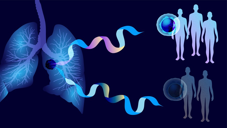
Neurological and neuropsychiatric disorders such as Parkinson’s disease, Alzheimer’s disease, and amyotrophic lateral sclerosis (ALS) are often diagnosed only after symptoms emerge. By that stage, underlying disease processes in the brain and nervous system may already be advanced. Because brain biopsy is limited by accessibility, there is growing interest in biomarkers that can be measured in biofluids. Building on this approach, researchers now report a framework for isolating neuron-specific extracellular vesicles from blood and cerebrospinal fluid.
The Wyss Institute at Harvard University developed the approach in collaboration with Brigham and Women’s Hospital, the Broad Institute of MIT and Harvard, and Harvard Medical School. The work focuses on extracellular vesicles, membrane-enclosed particles released by cells into biofluids that carry RNA transcripts and proteins. Because neuron-specific extracellular vesicles make up only a very small fraction of vesicles in plasma and other biofluids, the researchers developed methods to identify surface markers that could enable antibody-based isolation.
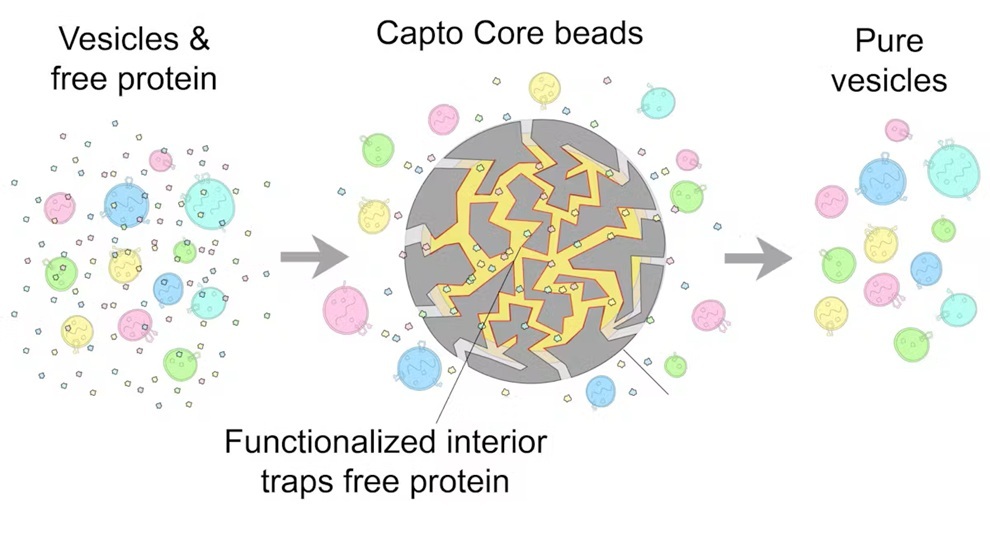
The team first established a method for isolating the full spectrum of extracellular vesicles from plasma and other body fluids at high purity. The technique uses porous beads that trap and remove free proteins such as albumin while excluding larger extracellular vesicles. According to the researchers, this one-step procedure increased extracellular vesicle purity by an additional 100-fold.
Using mass spectrometry, the researchers then generated reference proteomes for extracellular vesicles from plasma and cerebrospinal fluid. They combined these data with computational analysis of single-cell RNA sequencing studies to prioritize predicted transmembrane proteins whose genes were selectively expressed in neurons. One candidate marker, NRXN3, was confirmed on extracellular vesicles from both plasma and cerebrospinal fluid using ultrasensitive SIMOA detection assays and single-vesicle imaging methods.
The proof-of-concept study was published in Nature Neuroscience on October 23, 2026. The team also developed and validated an immuno-isolation method that reliably isolated NRXN3-carrying extracellular vesicles from human cerebrospinal fluid and plasma samples. The findings provide a framework for further analysis of proteins contained within neuron-derived extracellular vesicles and for identifying additional extracellular vesicle markers associated with other cell types and tissues.
“This project is a significant step towards our goal of reading out the brains of patients through the contents of neuron-specific EVs. We are now measuring specific proteins inside of these EVs to confirm their neuronal origin. Our goal is to look inside neuron-derived EVs for markers that are correlated with earlier stages and the progression of neurological disorders, as well as therapeutic efficacy of present and future drug candidates. More fundamentally, such markers can also help us better understand some of the underlying early pathologies, which for many neurological disorders are not clear at all,” said David Walt, Core Faculty member at the Wyss Institute.
Related Links
Wyss Institute at Harvard University