New AI Model Maps Where Glioblastoma Could Return After Surgery
Posted on 29 Sep 2026
Glioblastoma is the most common malignant brain tumor in adults and the most lethal, with median survival of about 17 months after diagnosis. Even after surgeons remove all visible tumor and patients receive additional treatment, the cancer almost always returns, typically at or near the surgical cavity. Being able to identify these high-risk areas before recurrence becomes visible on MRI could help guide treatment decisions.
Researchers at the University of California, San Francisco (UCSF) and the University of Michigan developed the approach using stimulated Raman histology (SRH) and FastGlioma, an AI system that scores tissue for tumor infiltration. SRH produces microscopic images of fresh, unprocessed tissue in less than a minute. Unlike SRH, conventional pathology requires processing tissue with dyes and stains, which takes longer and is more labor-intensive.
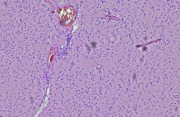
The researchers developed the model using about 300 tissue samples collected during glioblastoma surgery from 60 UCSF Health patients. They tested it separately on about 100 samples from another 20 patients. Median time to recurrence among the patients was 5.5 months. The team focused on first recurrence because experimental treatments given later in the disease can affect tumor growth and make subsequent recurrences harder to predict reliably.
The FastGlioma infiltration score alone predicted recurrence sites about as well as conventional pathology. The researchers then combined the score with clinical, imaging, and molecular data tested across six machine-learning models. The best-performing model was significantly more likely to distinguish sites that would develop recurrence from those that would not. Tumor infiltration was the strongest individual predictor in five of the six models, providing more predictive information than the tumor’s molecular characteristics. The model also performed well in predicting recurrence within 5 or 10 millimeters of sampled tissue.
The findings were published September 25, 2026, in Science Advances. The researchers said the predictions could inform decisions about removing additional tissue during surgery when safe, or targeting predicted sites with higher-dose focal radiation or drugs infused directly into the tumor. Their stated goal is to delay the first recurrence.
“The system has incredible potential to provide neurosurgeons with valuable real-time guidance during tumor removal. It can also generate insights that guide subsequent treatment decisions,” said Sanjeev Herr, MD, a postdoctoral research fellow at UC San Francisco and Drexel University College of Medicine.
Related Links
UCSF Health
University of Michigan Medical School