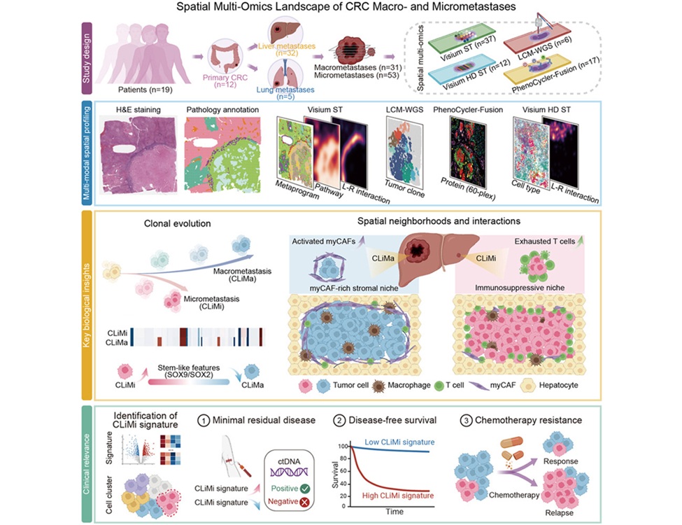
Mosaic Confocal Microscopy Technique Speeds Up Skin Cancer Surgery
By LabMedica International staff writers
Posted on 12 Feb 2014
A new and faster optical approach called strip mosaicing confocal microscopy was recently developed to reduce the time required to perform Mohs surgery for the removal of malignant skin cancers.Posted on 12 Feb 2014
Mohs surgery, also called Mohs micrographic surgery, is a precise surgical technique that is used to remove all parts of cancerous skin tumors while preserving as much healthy tissue as possible. Mohs surgery is used to treat such skin cancers as basal cell and squamous cell carcinomas.
.")
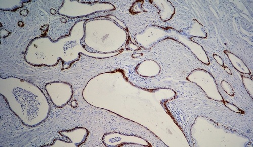
Image: Comparison of residual cancer detected with the new confocal imaging technique and the currently used freezing and staining technique (Photo courtesy of Dr. Milind Rajadyhyaksha, Memorial Sloan-Kettering Cancer Center).
Investigators at Memorial Sloan Kettering Cancer Center (New York, NY, USA) were funded by a grant from the [US] National Institute of Biomedical Imaging and Bioengineering (Bethesda, MD, USA) to develop a microscopy method to rapidly analyze tissues during the Mohs procedure.
The investigators developed a new pathological assessment technique called strip mosaicing confocal microscopy that employed a focused laser line to perform multiple scans of tissue excised during Mohs surgery to obtain image “strips” that were then combined, like a mosaic, into a complete image of the tissue. The process required only 90 seconds and eliminated the need to freeze and stain the tissue samples for analysis— a process that takes 20 to 45 minutes.
In a study, tissue samples from 17 Mohs cases were imaged in the form of strip mosaics. Each mosaic was divided into two halves (submosaics) and graded by a Mohs surgeon and a dermatologist who were blinded to the pathology. The 34 submosaics were compared with the corresponding Mohs pathology. Results revealed that the overall image quality was excellent for resolution, contrast, and stitching. Components of normal skin including the epidermis, dermis, dermal appendages, and subcutaneous tissue were easily visualized. The preliminary measures of sensitivity and specificity were both 94% for detecting skin cancer margins.
Dr. Steve Krosnick, director of the program for image-guided interventions at the [US] National Institute of Biomedical Imaging and Bioengineering, said, “The technology is particularly well-suited for Mohs-trained surgeons, who are experts at performing excisions and interpreting images of tissue samples removed during the Mohs procedure. Image quality, ability to make accurate interpretations, and time savings will be key parameters for adoption of the system in the clinical setting, and the current results are very encouraging.”
The study was published in the October 2013 issue of the British Journal of Dermatology.
Related Links:
Memorial Sloan Kettering Cancer Center
National Institute of Biomedical Imaging and Bioengineering

Gold Member
Nucleic Acid Extractor System
NEOS-96 XT


Rapid Sepsis Test
SeptiCyte RAPID

Immunofluorescence Analyzer
IFA System