Abdominal Fluid Testing Can Predict Ovarian Cancer Progression
Posted on 11 Feb 2026
Ovarian cancer kills more women than any other gynecological cancer, largely because it is usually diagnosed only after it has spread widely within the abdomen. Unlike many other cancers, it does not rely on blood vessels to metastasize, making its rapid progression difficult to track and predict. Researchers have now uncovered a key mechanism that explains how ovarian cancer advances so aggressively and resists treatment, opening up new diagnostic and therapy possibilities.
In a study led by Nagoya University (Nagoya, Japan), researchers investigated how ovarian cancer spreads within the abdominal cavity. By analyzing abdominal fluid from patients, the team discovered that cancer cells do not act alone during dissemination. Instead, they recruit mesothelial cells, which normally line and protect the abdominal cavity, to form cooperative hybrid clusters.
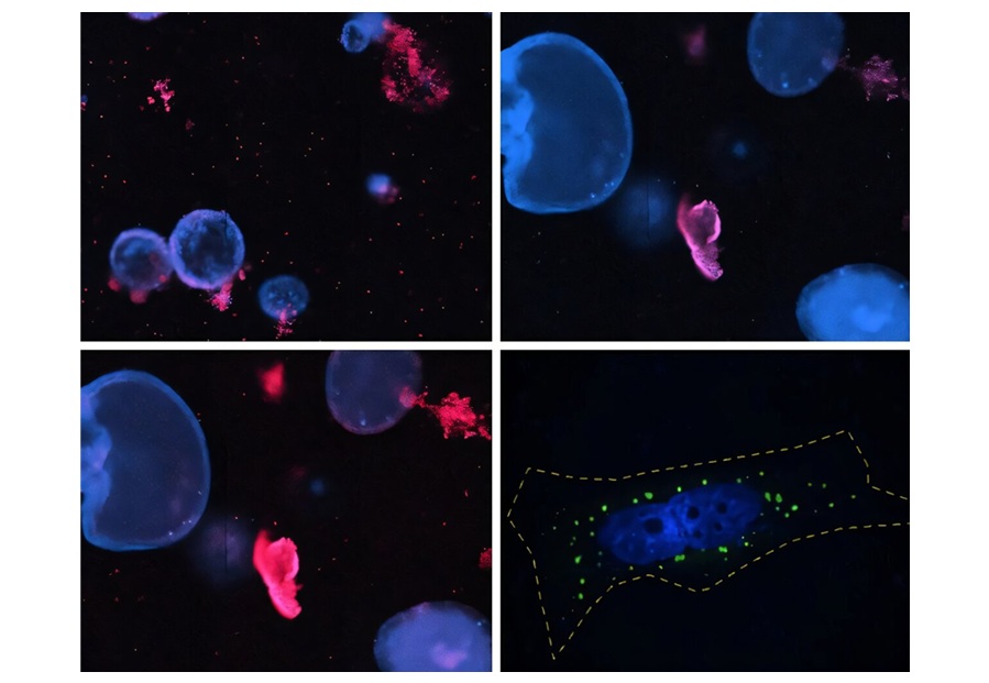
 stick to mesothelial cells (green) and form hybrid spheres that cut into surrounding abdominal tissue (Photo courtesy of Uno et al., 2026)")
Cancer cells release the signaling protein TGF-β1, which transforms mesothelial cells and induces the formation of invadopodia—spike-like structures capable of cutting through tissue. These mesothelial cells lead the invasion process, creating physical pathways that cancer cells then follow. This strategy allows cancer cells to spread efficiently without undergoing major genetic changes themselves.
Using advanced microscopy, mouse models, and single-cell genetic analysis, the researchers observed that about 60% of ovarian cancer cell clusters contained recruited mesothelial cells. These hybrid spheres were significantly more invasive and showed greater resistance to chemotherapy than cancer cells alone.
The discovery, published in Science Advances, reveals a previously hidden stage of ovarian cancer spread, occurring while cells float freely in abdominal fluid before attaching to new organs. Targeting the interaction between cancer cells and mesothelial accomplices—such as blocking TGF-β1 signaling—could open new therapeutic avenues. Monitoring these hybrid cell clusters in abdominal fluid may also help predict disease progression and treatment response.
Related Links:
Nagoya University