Ultra-Rapid Genetic Test Diagnoses Brain Tumors in Two Hours
Posted on 21 May 2025
The standard process for treating brain tumors typically begins with an MRI scan to detect the presence of a tumor. Afterward, patients meet with clinicians to discuss potential tumor types. For many tumor cases, the next step is surgery to obtain a sample from the tumor, which is then sent to centralized labs for testing to examine any DNA abnormalities. The purpose of this test is to determine the tumor’s classification. Historically, experts have analyzed the specimens visually through neuropathology, trying to identify the cells under a microscope.
However, in recent years, this method has changed, and tumors are now categorized based on genetic and DNA abnormalities. This change, although a significant advancement, has led to delays due to technological constraints. It often takes 6-8 weeks or more to receive the complete results, which inform patients about their tumor type and prognosis. This waiting period is distressing for patients, and it also postpones the initiation of treatments such as radiotherapy and chemotherapy, which may decrease their effectiveness. Now, a groundbreaking ultra-rapid method for genetically diagnosing brain tumors could significantly reduce this waiting time. The new method is fast enough to provide results within just a couple of hours, potentially allowing this crucial information to be available during surgery, thus informing surgical decisions in real-time.
")
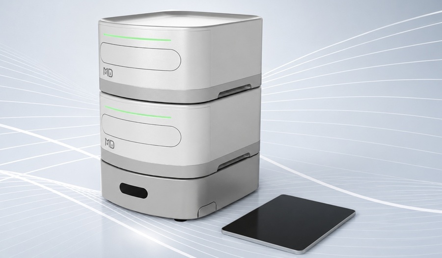
This innovative technique, developed by researchers at the University of Nottingham (Nottingham, UK), dramatically reduces the time needed to classify brain tumors, cutting the process from 6-8 weeks down to as little as two hours, improving patient care. The team developed a method to sequence specific regions of human DNA at higher depths, using portable sequencing devices from Oxford Nanopore Technologies (Oxford, UK). This method allows for a much quicker examination of relevant parts of the human genome and enables the simultaneous sequencing of multiple DNA regions, speeding up the overall process. Their method uses ROBIN, a software tool that operates on the P2 PromethION nanopore sequencers. This technology sequences DNA by detecting changes in current flow as single DNA molecules pass through a nanopore, a tiny hole in a membrane.
Once a tissue sample is removed during surgery, it is sent to the pathology lab, where the DNA is extracted and forwarded to the research team for sequencing. The team tested this method on brain tumor samples, using it in 50 surgeries as described in their study published in Neuro-Oncology. The results were exceptional, with the approach achieving a 100% success rate in providing rapid intraoperative diagnoses. Within two hours of surgery, the method delivered diagnostic results and tumor classifications in just minutes. Additionally, the sequencing platform’s continuous operation allows for a fully integrated diagnosis to be completed within 24 hours.
“Traditionally, the process of diagnosing brain tumors has been slow and expensive. Now, with this new technology we can do more for patients because we can get answers so much more quickly which will have a much bigger influence on clinical decision making, in as little as two hours. Patients find waiting many weeks for results extremely difficult and this adds to the anxiety and worry at what is already a very difficult time,” said Dr. Stuart Smith, a neurosurgeon from the School of Medicine at the University of Nottingham. “This type of operation can be quite long, so potentially, a surgeon could be informed during surgery of the accurate diagnosis, which would then impact on the surgical strategy.”
Related Links:
University of Nottingham
Oxford Nanopore Technologies