Elevated Plasma Levels of Glial Fibrillary Acidic Protein Indicate Increased Alzheimer’s Risk
By LabMedica International staff writers
Posted on 01 Mar 2021
A recent study has demonstrated that plasma levels of the protein GFAP (glial fibrillary acidic protein) are elevated in cognitively normal older adults at risk of developing Alzheimer’s disease.Posted on 01 Mar 2021
GFAP is a protein in the cytoskeleton of brain astrocytes. Previous studies have shown that it can be measured in blood samples and is associated with Alzheimer’s disease (AD). However, plasma GFAP has not been investigated in cognitively normal older adults at risk of AD, based on brain amyloid-beta (Abeta) load.
")
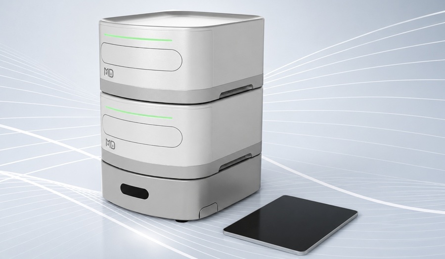
Image: The HD-X Analyzer is the latest model of the fully automated Simoa bead-based immunoassay platform. (Photo courtesy of Quanterix)
In the current study, investigators at Edith Cowan University (Perth, Australia) compared plasma GFAP levels between cognitively normal older adults with low brain Abeta load (Abeta−) and cognitively normal older adults at risk of AD, due to high brain Abeta load, (Abeta+). They postulated that plasma GFAP levels would be higher in the Abeta+ group compared to the Abeta− group.
To confirm this hypothesis, the investigators used the Quanterix (Billerica, MA, USA) Simoa GFAP Discovery Kit on the ultra-sensitive Single molecule array (Simoa) platform (HDx instrument) to measure levels of GFAP, Abeta1–42, and Abeta1–40. Cross-sectional analyses were carried out for plasma GFAP and plasma Abeta1–42/Abeta1–40 ratio, a blood-based marker associated with brain Abeta load, in participants (65–90 years) categorized into low (Abeta−) and high (Abeta+) brain Abeta load groups via Abeta positron emission tomography (PET).
Results revealed that plasma GFAP levels were significantly higher, and plasma Abeta1–42/Abeta1–40 ratios were significantly lower, in Aβ+ participants compared to Aβ− participants. This finding demonstrated that plasma GFAP levels were elevated in cognitively normal older adults at risk of AD. Furthermore, these observations suggested that astrocytic damage or activation began from the pre-symptomatic stage of AD and was associated with brain Abeta load.
"Blood biomarkers are becoming an exciting alternative to the existing expensive and invasive methods of diagnosing Alzheimer's disease," said senior author Dr. Ralph N. Martins, professor of aging & Alzheimers disease at Edith Cowan University. "The GFAP biomarker could be used to develop a simple and quick blood test to detect if a person is at very high risk of developing Alzheimer's. Early diagnosis is critical to allow us to implement medication and lifestyle interventions that can help delay the progression of the disease and give people more time before symptoms develop."
The study was published in the January 11, 2021, online edition of the journal Translational Psychiatry.
Related Links:
Edith Cowan University
Quanterix
.jpg)
Gold Member
Clinical Chemistry Assay
Sorbitol Dehydrogenase (SDH)


CMV CLIA Diagnostic
CLIA CMV IgA Screen Group

Automated Clinical Chemistry Analyzer
Envoy 500+