Biomarker in Stools Helps to Diagnose Necrotizing Enterocolitis in Premature Babies
By LabMedica International staff writers
Posted on 20 Nov 2019
Identification of a novel biomarker will enable clinicians to better diagnose necrotizing enterocolitis, a devastating disease that affects premature infants.Posted on 20 Nov 2019
Necrotizing enterocolitis (NEC) is a medical condition where a portion of the bowel dies. It typically occurs in newborns of either sex that are either premature or otherwise unwell. Symptoms may include poor feeding, bloating, decreased activity, blood in the stool, or vomiting of bile. Inflammation of the intestine leads to bacterial invasion causing cellular damage and cell death, which causes necrosis of the colon and intestine. About 7% of those that are born premature develop NEC. Onset is typically in the first four weeks of life. Among those affected, about 25% die.

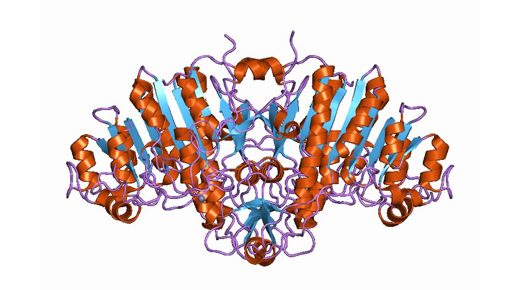
Image: Cartoon representation of the molecular structure of alkaline phosphatase protein (Photo courtesy of Wikimedia Commons)
Since intestinal alkaline phosphatase (iAP) activity is known to signal the chemical process triggering inflammation, investigators at Louisiana State University Health Sciences Center (New Orleans, USA) examined the abundance and enzyme activity of iAP shed in stools by newborns to assess the correlation of two iAP biochemical measures with NEC disease severity.
Intestinal alkaline phosphatase is secreted by enterocytes, and seems to play a pivotal role in intestinal homeostasis and protection as well as in mediation of inflammation via repression of the downstream Toll-like receptor (TLR)-4-dependent and MyD88-dependent inflammatory cascade. It dephosphorylates toxic/inflammatory microbial ligands like lipopolysaccharides, unmethylated cytosine-guanine dinucleotides, flagellin, and extracellular nucleotides such as uridine diphosphate or ATP. Thus, altered IAP expression has been implicated in chronic inflammatory diseases such as inflammatory bowel disease (IBD). It also seems to regulate lipid absorption and bicarbonate secretionin the duodenal mucosa, which regulates the surface pH.
The current multi-center diagnostic study comprised 136 premature infants (gestational age, less than 37 weeks). Infant stool samples were collected between 24 and 40 or more weeks post-conceptual age. Enrolled infants underwent abdominal radiography at physician and hospital site discretion. Enzyme activity and relative abundance of iAP were measured using fluorometric detection and immunoassays, respectively.
The data showed that of the 136 infants, 68 (50.0%) were male, median birth weight was 1050 g, and median gestational age was 28.4 weeks. A total of 25 infants (18.4%) were diagnosed with severe NEC, 19 (14.0%) were suspected of having NEC, and 92 (66.9%) did not have NEC; 26 patients (19.1%) were diagnosed with late-onset sepsis, and 14 (10.3%) had other non–gastrointestinal tract infections.
Results of iAP analysis revealed that high amounts of intestinal alkaline phosphatase protein in stools combined with low intestinal alkaline phosphatase enzyme activity were associated with diagnosis of necrotizing enterocolitis. There was no association of intestinal alkaline phosphatase levels with non–gastrointestinal tract infections.
"Intestinal AP is the first candidate diagnostic biomarker, unique in its predictive value for NEC," said senior author Dr. Sunyoung Kim, professor of biochemistry and molecular biology at Louisiana State University Health Sciences Center. "It is correlated only with NEC and is not associated with sepsis or other non-GI infections. The clinical potential of this noninvasive tool lies in its use to identify infants most at risk to develop NEC, to facilitate management of feeding and antibiotic regimens, and monitor response to treatment."
The study was published in the November 8, 2019, online edition of the journal JAMA Network Open.
Related Links:
Louisiana State University Health Sciences Center


