Novel Non-Invasive Sampling Technique Diagnoses Cutaneous Leishmaniasis
By LabMedica International staff writers
Posted on 27 Jul 2017
The definitive diagnosis of cutaneous leishmaniasis requires parasitological detection and it is usually based on direct detection of amastigotes in smears from lesions by microscopy and by the culturing of promastigotes from infected tissues.Posted on 27 Jul 2017
Although these techniques are highly specific, they require highly skilled health workers and have the inherent risks of all invasive procedures, such as pain and risk of bacterial and fungal super-infection. Therefore, it is essential to reduce discomfort, potential infection and scarring caused by invasive diagnostic approaches especially for children.
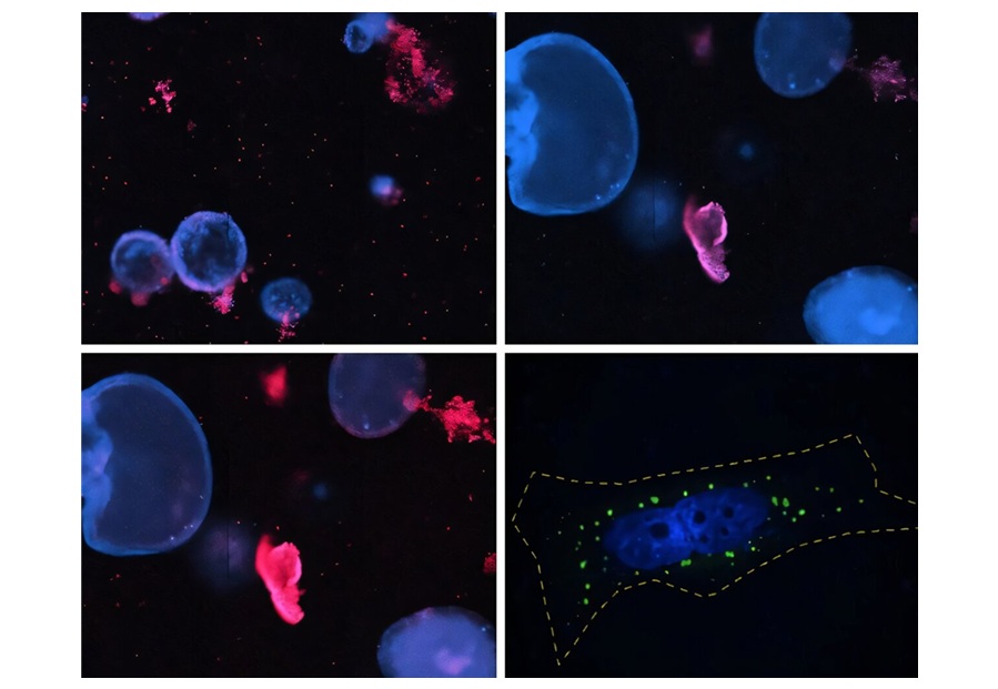
 Determination of the lesion for disc sampling; B) Application of the tape strip disc on the lesion; C) Even pressure applied for 20 seconds with the plunger; and D) Detaching the disc and transferred into a sterile vial for DNA isolation (Photo courtesy of the Pasteur Institute of Iran).")
Image: A) Determination of the lesion for disc sampling; B) Application of the tape strip disc on the lesion; C) Even pressure applied for 20 seconds with the plunger; and D) Detaching the disc and transferred into a sterile vial for DNA isolation (Photo courtesy of the Pasteur Institute of Iran).
Scientists at the Pasteur Institute of Iran (Tehran, Iran) working with their colleagues at the Imperial College London (London, UK) recruited 119 patients suspected of suffering from cutaneous leishmaniasis with different clinical manifestations and samples were collected both from their lesions and from uninfected areas. In addition, 15 fungal-infected lesions and 54 areas of healthy skin were examined. There were 49 female and 70 males and the median age was 30 years (average 33 ± 20.3, range: 1 to 79 years old). The touch smear of dermal tissue of each patient was prepared by superficial incision on the margin of the lesion against a glass microscope slide, stained and examined.
For non-invasive skin sampling, one tape disc was placed on selected areas (infected and non-infected areas). In order to apply even pressure to the disc, a plunger was gently held on the selected areas and pressed for approximately 20 seconds. DNA was isolated from the strip and two polymerase chain reactions (PCR) assays were used in the study: First PCR was targeting the kDNA1 minicircle of Leishmania species and the second was for species identification (L. tropica and L. major).
The tape stripping technique used was child-friendly, causes virtually no pain, and is very practical for those lesions, which occur in sensitive areas such as the eyelids, lips and ears. Of the 119 confirmed CL patients enrolled in this study, 79 patients (66%) presented with acute CL (duration ranged from one up to 12 months), 32 (27%) with chronic CL (duration ranged from 30 up to 360 months). In total, 71 patients were infected with L. tropica (60%), 41 with L. major (34%) and seven were undetermined (6%). The highest diagnostic sensitivity of 100% was achieved with the tape strip disc technique. Of 31 suspected patients, only 16 were positive for culturing parasite from lesion biopsies (sensitivity 51%), which was significantly lower than tape strip disc sampling.
The authors concluded that the duration of sampling is short (less than one minute) and species identification by PCR is highly specific and sensitive. The sequential tape stripping sampling method is a sensitive, non-invasive and cost-effective alternative to traditional diagnostic assays and it is suitable for field studies as well as for use in health care centers. The study was published on July 13, 2017, in the journal Public Library of Science Neglected Tropical Diseases.
Related Links:
Pasteur Institute of Iran
Imperial College London

Gold Member
Quantitative POC Immunoassay Analyzer
EASY READER+


Food Allergy Screening ELISA Kit
Allerquant 14G B ELISA

Thyroid Test
Anti-Thyroid EIA Test