DNA-Barcode Profiling Identifies Cancer Biomarkers in Fine Needle Aspirates
By LabMedica International staff writers
Posted on 30 Jan 2014
A recent paper described a method that capitalizes on DNA-barcode-antibody sensing to enable analysis of a multitude of proteins obtained from minimally invasive fine-needle aspirates. Posted on 30 Jan 2014
Traditionally, immunohistochemistry-based clinical diagnoses have required invasive core biopsies and the use of a limited number of protein stains to identify and classify cancers. In a recent paper, investigators at the Massachusetts General Hospital (Boston, USA) introduced a technology that allows analysis of hundreds of proteins from minimally invasive fine-needle aspirates (FNAs), which contain much smaller numbers of cells than core biopsies.
.")
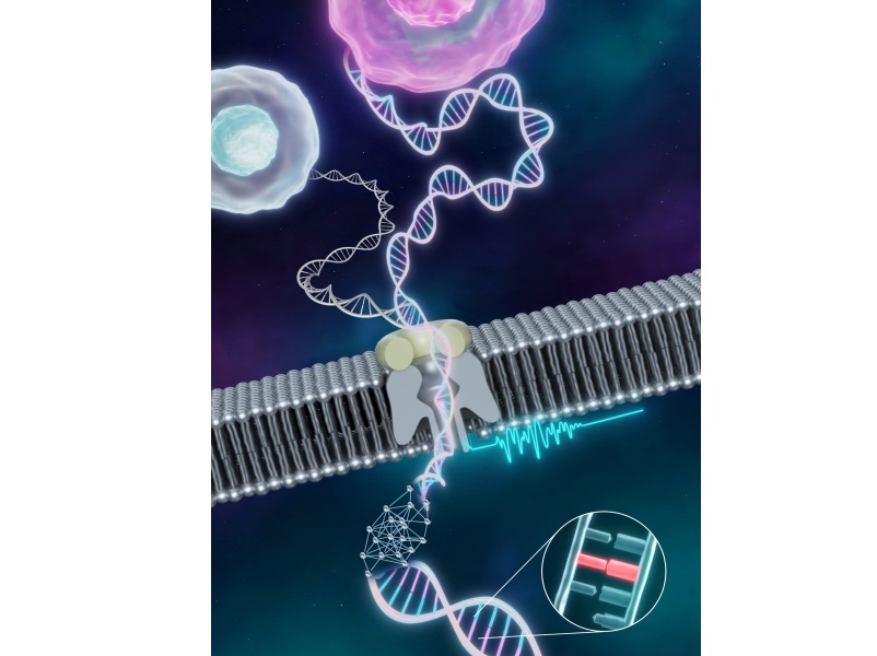
Image: A method based on DNA-barcode-antibody sensing allows analysis of a multitude of proteins from minimally invasive fine-needle aspirates (Photo DNA-barcode schematic, courtesy of Roche).
The method is based on DNA-barcode-antibody sensing, where the barcodes can be photocleaved and digitally detected without any amplification steps. When mixed with a tumor sample, the antibodies bind to their specific targets. A light pulse then releases the unique DNA-labeled bound antibodies, which are then tagged with fluorescently labeled, complementary DNA barcodes. The barcodes can be detected and quantified via imaging, revealing which markers were present in the sample.
This approach was applied to profile about 90 proteins in cells obtained from FNAs. The data was then used to map patient heterogeneity at the protein level. Results published in the January 15, 2014, issue of the journal Science Translational Medicine revealed that this method showed high reproducibility and could achieve single-cell sensitivity.
"What this study sought to achieve was to vastly expand the information that we can obtain from just a few cells," said senior author Dr. Cesar Castro, a researcher in hematology and oncology at the Massachusetts General Hospital. "Instead of trying to procure more tissue to study, we shrank the analysis process so that it could now be performed on a few cells. We showed that this technology works well beyond the highly regulated laboratory environment, extending into early-phase clinical trials. Ultimately, the implications for this type of technology could be vast. In this era of personalized medicine, we could leverage such technology not only to monitor but actually to predict treatment response. By obtaining samples from patients before initiating therapy and then exposing them to different chemotherapeutics or targeted therapies, we could select the most appropriate therapy for individual patients."
Related Links:
Massachusetts General Hospital
New
Gold Member
POC Helicobacter Pylori Test Kit
Hepy Urease Test


Food Allergy Screening ELISA Kit
Allerquant 14G B ELISA

Urine Analyzer
respons® UDS100