Melanoma Identified by Overexpression of Protein IMP-3
By Labmedica staff writers
Posted on 28 Jul 2008
The insulin-like growth factor-II mRNA binding protein, IMP-3, is produced excessively in malignant melanoma but not in harmless moles. This finding might give doctors a new, objective way to distinguish melanoma from some benign moles that look like melanoma but are not cancerous.Posted on 28 Jul 2008
Pathologists play a major role in diagnosing and staging skin cancers, by sorting through neoplasms and identifying features. They analyze cells within the lesions and apply chemical stains and other tools to measure the depth and predict future behavior of the growths. A pilot study showed why IMP-3 could become an important tool for pathologists.
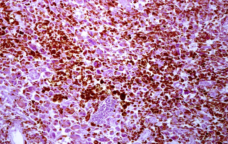
The study was performed by Jennifer G. Pryor, M.D., a resident from the University of Rochester Medical School (URMC; NY, USA) department of pathology and laboratory medicine, colleagues from the University's department of dermatology, and the Dako Corp. (Carpinteria, CA, USA). The researchers investigated samples of 56 biopsied lesions from 48 adults. The lesions fell into the category of cutaneous melanocytic neoplasms, a diverse group that includes benign moles; Spitz nevi, a type of mole seen in younger people that can be easily mistaken for melanoma but is not cancerous; and malignant melanoma, which has several phases of growth.
The investigators found that none of the benign moles or the benign moles with irregular features and some abnormal cells overexpressed the IMP-3 protein. However, the protein was produced excessively in most melanomas, and especially in metastatic melanomas. IMP-3 was also overexpressed in cases of invasive thin melanomas. This is significant because most thin melanomas have a good prognosis, but some act more aggressively and currently there is no accurate way to distinguish between the different types of thin lesions.
In previous studies, expression of the IMP-3 protein has been linked to pancreas, kidney, ovary, and lung cancers, but this is the first time a connection to melanoma has been demonstrated, according to Dr. Pryor. Additional work is needed to compare IMP-3 expression with long-term survival data from thin melanoma patients, to find out if patients whose tumors express IMP-3 might benefit from more careful monitoring and aggressive treatment, the study's investigators noted.
The study was published in the January 2008, edition of the journal Modern Pathology.
Related Links:
University of Rochester Medical School
Dako

Gold Member
Aspiration System
VACUSAFE


HPV Molecular Test
BD Onclarity HPV Assay

Thyroid Test
Anti-Thyroid EIA Test