Powassan Virus Diagnosed in Patients with Tick-Borne Diseases
By LabMedica International staff writers
Posted on 02 Aug 2017
Powassan virus (POWV) lineage II, also known as deer tick virus, is an emerging tick-borne flavivirus transmitted by Ixodes scapularis ticks, which are also the primary vector for the Lyme disease pathogen Borrelia burgdorferi.Posted on 02 Aug 2017
The seroprevalence of POWV in humans in some regions of North America is known with a range of 0.5% to 3.3%, but because the geographic distribution is quite extensive, the seroprevalence of most at-risk populations is uncertain. POWV is typically detected with an immunoglobulin M (IgM) antibody capture e enzyme-linked immunosorbent assay (ELISA) or an IgM immunofluorescence antibody (IFA) assay.

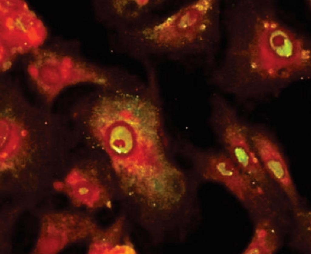
Image: Immunofluorescent antibody stain showing positive Powassan virus-infected cells (Photo courtesy of Coppe Laboratories).
Scientists at the Marshfield Clinic Research Foundation (Minocqua, WI, USA) and their colleagues selected 95patients with suspected tick-borne disease (TBD) and 50 patients undergoing routine chemical screening who sought treatment during July to August 2015 at the Marshfield Clinic in northern Wisconsin, a TBD-endemic area. Patients were considered to have suspected TBD if a serologic test for B. burgdorferi was ordered.
The investigators performed screening assays on all specimens for tick-borne encephalitis virus complex (TBEV-C) and B. burgdorferi and performed POWV serology on TBEV-C–positive specimens. They evaluated heterologous flavivirus cross-reactivity, by performing the West Nile virus (WNV) enzyme immunoassay with TBEV-C–positive samples. They also performed the EUROIMMUN Flavivirus Mosaic Panel, an IgG IFA assay panel including tests for TBEV, WNV, yellow fever virus, dengue viruses 1–4, and Japanese encephalitis virus, on samples positive for POWV IgG by the IFA assay.
Serologic evidence of POWV infection was present in nine (9.5%) TBD patients and two (4.0%) patients with routine chemistry screening completed. Similar to other flavivirus serologic assays, considerable cross-reactivity occurred with the Flavivirus Mosaic IgG IFA assay. The fluorescence intensity was stronger for TBEV than it was for other flaviviruses in all TBD patients except for one patient with prior confirmed WNV infection. Evidence of current or prior B. burgdorferi infection was present in 63 (66.3%) TBD patients. Of the 41 (43.2%) TBD patients with evidence of B. burgdorferi infection, seven (17.1%) had serologic evidence of acute POWV infection and three (7.3%) had laboratory-confirmed POWV infection.
The authors concluded that in a Lyme disease–endemic area, POWV seroreactivity and confirmed POWV infection were present. The spectrum of disease is broader than previously realized, with most patients having minimally symptomatic infection. The study was published in the August 2017 issue of the journal Emerging Infectious Diseases.
Related Links:
Marshfield Clinic Research Foundation


