Enterovirus Infections Linked with Autoimmunity Leading to Diabetes
By LabMedica International staff writers
Posted on 02 Feb 2017
Type 1 diabetes is caused by an immune-mediated process that damages insulin-producing beta cells in the pancreas and the subclinical phase of the disease can be identified by detecting autoantibodies.Posted on 02 Feb 2017
Enteroviruses have been linked to type 1 diabetes (T1D) in studies showing an increased frequency of these viruses in the blood and pancreas of diabetic and autoantibody-positive individuals and in studies showing an increased frequency of enterovirus antibodies in people with T1D.
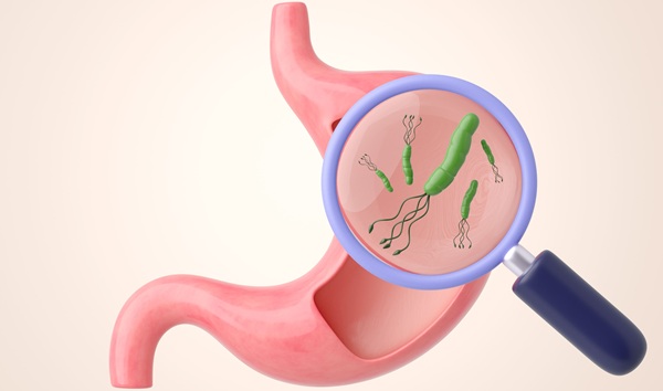
 revealed the presence of coxsackievirus virus particles, which were found within a specimen of muscle tissue (Photo courtesy of Dr. Fred Murphy and Sylvia Whitfield).")
Image: A transmission electron photomicrograph (TEM) revealed the presence of coxsackievirus virus particles, which were found within a specimen of muscle tissue (Photo courtesy of Dr. Fred Murphy and Sylvia Whitfield).
Virologists at the University of Tampere and their colleagues analyzed whether the presence of enteroviruses in stools was associated with the appearance of islet autoimmunity in the "Type 1 Diabetes Prediction and Prevention Study" in Finland. Altogether, 1,673 longitudinal stool samples from 129 case children who turned positive for multiple islet autoantibodies and 3,108 stool samples from 282 matched control children were screened for the presence of enterovirus ribonucleic acid RNA using polymerase chain reaction (RT-PCR). Viral genotype was detected by sequencing.
The team diagnosed 108 infections in the 129 case children and 169 infections in the 282 control children during the whole follow-up (mean 0.8 versus 0.6 infections per child). This difference was also seen in infections that occurred prior to the appearance of autoantibodies (0.6 versus 0.4 infections per child). Further analyses showed that the excess of infections in case children occurred more than 12 months before the first autoantibody-positive sample was taken. During this time period, an average of 0.62 infections were diagnosed per case child compared with 0.33 infections per control child, corresponding to 6.3 versus 2.1 infections per 10 follow-up years. The most frequent enterovirus types included coxsackievirus A4 (28% of genotyped viruses), coxsackievirus A2 (14%) and coxsackievirus A16 (11%).
The authors concluded their present study suggests that enterovirus infections in young children are associated with the appearance of islet autoantibodies with a time lag of about one year. This finding supports previous observations from other prospective studies suggesting that enterovirus infections may play a role in the initiation of the beta cell-damaging process. The study was published on January 9, 2017, in the journal Diabetologia.

Gold Member
Automatic Hematology Analyzer
CF9600


Rapid Sepsis Test
SeptiCyte RAPID

Automatic CLIA Analyzer
Shine i6000