Non-Hodgkin Lymphoma Linked to Q Fever Bacterium
By LabMedica International staff writers
Posted on 26 Oct 2015
The incidence of non-Hodgkin lymphoma (NHL), associated with significant mortality, is increasing in many regions, making it a global health challenge for the coming years.Posted on 26 Oct 2015
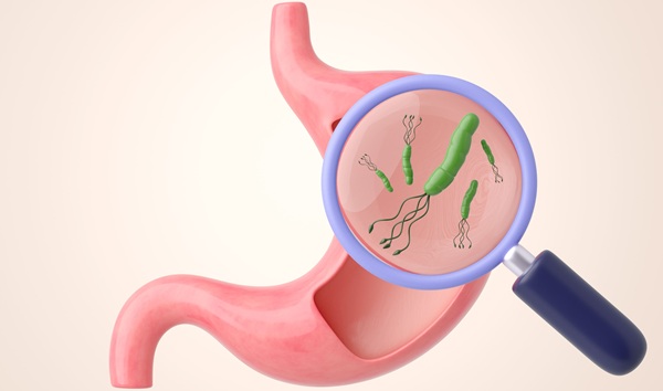
The bacterium Coxiella burnetii causes Q fever, which is an infectious disease that is primarily transmitted through the excrement of cattle, sheep, and goats and humans contract it from these animals is associated with an increased risk of lymphoma.
.")
Image: A dry fracture of a Vero cell exposing the contents of a vacuole where Coxiella burnetii, the causative agent of Q fever, are rapidly growing (Photo courtesy of National Institutes of Health).
A large team of French scientists led by those at the Aix-Marseille University (Marseille, France) screened 1,468 patients treated at the French National Referral Center for Q fever from 2004 to 2014. Acute Q fever was defined by the association of clinical symptoms (fever, hepatitis and/or pneumonia) with the serological criteria of a phase II immunoglobulin G (IgG) titer equal to or greater than 200 and a phase II IgM titer equal to or greater than 50, seroconversion or a positive polymerase chain reaction (PCR) and no endocarditis.
Formalin-fixed and paraffin-embedded biopsy samples were analyzed by an expert hematopathologist in all cases to confirm the diagnosis of lymphoma. Immunofluorescence (IF), 16S ribosomal ribonucleic acid (rRNA) fluorescence in situ hybridization (FISH) and genomic DNA FISH were performed as well as other diagnostic techniques. The amount of interleukin-10 (IL-10) in sera was determined using specific immunoassays (R&D Systems; Minneapolis, MN, USA). IL10 and Tumor necrosis factors (TNF) were measured in supernatants from non-stimulated and C. burnetii stimulated peripheral blood mononuclear cells (PBMC).
The team found, based on their analysis, that patients with Q fever were 25 times more likely to develop diffuse large B-cell lymphoma than those without the infection. In addition, the odds of lymphoma in patients with persistent concentrated infections are higher than those with other forms of Q fever. The investigators observed that Q fever patients with lymphoma demonstrated overproduction of the critical anti-inflammatory pathway IL-10, suggesting that suppression of the immune system may have allowed the lymphoma cells to evade immune detection and multiply.
Didier Raoult, MD, PhD, the senior author of the study, said, “As we continue to learn more about the association between C. burnetii and lymphoma, these results should encourage clinicians to survey high-risk patients as early as possible for potential cancer. Ultimately, this early diagnosis and treatment would improve outcomes for Q fever patients who subsequently develop lymphoma, particularly those with B-cell non-Hodgkin lymphoma.” The study was published on October 13, 2015, in the journal Blood .
Related Links:
Aix-Marseille University
R&D Systems

Gold Member
Aspiration System
VACUSAFE


Benchtop Thermomixer
Biometra TS1 ThermoShaker

Manual Pipetting Aid
Pipette Controllers macro