Lateral Flow Immunoassay Developed for Melioidosis
By LabMedica International staff writers
Posted on 03 Apr 2014
A lateral flow immunoassay that can be used in the clinical setting to diagnose melioidosis in 15 minutes has been developed and the test promises to provide improved management of patients with the disease. Posted on 03 Apr 2014
Isolation of Burkholderia pseudomallei, a soil-dwelling bacterium and the causative agent of melioidosis, from clinical samples is the “gold standard” for the diagnosis of melioidosis, but this can take as long as three days to a week to produce the results.

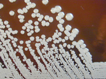
Image: The Gram-negative aerobic bacteria Burkholderia pseudomallei grown on sheep blood agar for 96 hours (Photo courtesy of Larry Stauffer).
Scientists at InBios International, Inc., (Seattle, WA, USA) and a team of international colleagues, have developed a rapid point-of-care antigen detection assay for the diagnosis of melioidosis. A diagnostic immunoassay based on the detection of B. pseudomallei capsular polysaccharide (CPS) was investigated. Following production of a CPS-specific monoclonal antibody (mAb), an antigen-capture immunoassay was developed to determine the concentration of CPS within a panel of melioidosis patient serum and urine samples.
The same mAb was used to produce a prototype Active Melioidosis Detect Lateral Flow Immunoassay (AMD LFI) and the limit of detection of the LFI for CPS is comparable to the antigen-capture immunoassay, approximately 0.2 ng/mL. LFIs were read after 15 minutes and determined to be positive or negative based on the presence or absence of a pink-red line at the test line in the presence of a positive control line. The analytical reactivity or inclusivity of the AMD LFI was 98.7% (76/77) when tested against a large panel of B. pseudomallei isolates. Analytical specificity or cross-reactivity testing determined that 97.2% of B. pseudomallei near neighbor species (35/36) were not reactive.
The authors concluded that when the LFI is used to test urine, sputum, and pus, high sensitivity will be achieved due to the increased colony forming units (CFU)/mL values in these matrices. The LFI is currently being evaluated in Thailand and Australia and the focus is to optimize and validate testing procedures on melioidosis patient samples prior to initiation of a large, multisite preclinical evaluation. The study was published on March 20, 2014, in the journal Public Library of Science Neglected Tropical Diseases.
Related Links:
InBios International Inc.


