Single Virus Detection Techniques Diagnoses Diseases Faster
By LabMedica International staff writers
Posted on 20 Jun 2013
Optics-based methods have been developed for determining the exact viral load of a sample by counting individual virus particles.Posted on 20 Jun 2013
The viral load is a measurement of the severity of a viral infection, and helps clinicians try to gauge how many viruses are packed into a certain volume of blood or other bodily fluid.
.")
Image: The LUCAS Holographic Microscope (Photo courtesy of Holomic LLC).
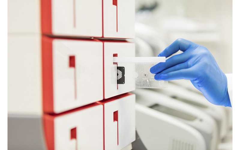
Two groups of scientists and engineers at the University of California campuses in Los Angles (UCLA; CA, USA) and Santa Cruz (UCSC; CA, USA) collaborated to develop methods that are faster and cheaper than standard tests and they offer the potential to conduct the measurements in a medical office or hospital instead of a laboratory. The bioengineers at UCLA are working to directly image single virus particles using holographic microscopy, while the team at UCSC is detecting single particles tagged with fluorescent labels on a microfluidic chip.
The UCLA team has demonstrated the ability to capture optical images of single viruses and nanoparticles over a comparatively large field of view using nanolenses that self-assemble around the virus particles like little magnifying glasses. This wide field of view allows the device to form images of many nanoparticles in a single photograph and provides a high-throughput platform for a direct and accurate viral load count. The instrument can be made sufficiently compact and lightweight for field applications and, attached to a cell phone, could become useful even in remote locations.
The UCSC team counts the viruses by detecting their nucleic acids, which is the genetic makeup of the viruses. The nucleic acids are labeled with a fluorescent dye, and light from the fluorescence is detected as they pass through a channel in a microfluidic chip about the size of a thumbnail. The labeling technique would allow clinicians to target specific viruses while ignoring unlabeled background material. This makes the process potentially useful in situations where clinicians already know what they are looking for, which is often the case for viral load tests.
Aydogan Ozcan, PhD, the lead scientist from UCLA, said, “Because viruses are very small compared to the wavelength of light, conventional light microscopy has difficulty producing an image due to weak scattering of subwavelength particles.” The team's new nanolens-nanoparticle assembly projects a hologram that can be recorded using a complementary metal-oxide-semiconductor (CMOS) imager chip, and digitally reconstructed to form an optical image of the particle. The study was presented at the Conference on Lasers and Electro-Optics held June 9–14, 2013, in San Jose (CA, USA).
Related Links:
University of California Los Angeles
University of California Santa Cruz

Gold Member
Automatic Hematology Analyzer
CF9600


Manual Pipetting Aid
Pipette Controllers macro

Automated Clinical Chemistry Analyzer
Envoy 500+