


Single-Vesicle Analysis Reveals Tissue-Associated Inflammatory Signatures
Posted on 07 Nov 2026
Chronic inflammation can persist for months or years when immune cells continue attacking tissues despite the absence of an active threat. It is associated with cancers, brain disorders, autoimmune diseases, and heart disease, and prolonged inflammation can lead to irreversible tissue damage. Conventional biomarkers can detect inflammation but may not indicate where it is occurring in the body. To provide more localized insight, researchers have developed a method for analyzing single extracellular vesicles to identify tissue-associated inflammatory signatures.
Nagoya University researchers developed a cellulose nanofiber (CNF) sheet-based technique that analyzes information on the surface and inside individual extracellular vesicles (EVs) in blood and urine. The method is designed to connect internal microRNA signals with membrane proteins without disrupting the vesicle membrane. This preserves the relationship between molecular cargo and tissue-associated surface markers, enabling detection of EV subpopulations that may otherwise be obscured when vesicles are broken open.
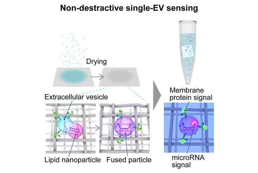
The CNF sheets are made from wood-derived cellulose and contain mesh-like nanoscale pores that capture and retain EVs. During drying, the sheets bring membrane-bound particles into close contact and promote membrane fusion. The researchers used this effect to fuse EVs with lipid nanoparticles carrying molecular beacons that fluoresce when they bind specific microRNAs.
To test the approach, the team created a laboratory model of chronic active antibody-mediated rejection, a complication of kidney transplantation, by exposing human kidney cells to inflammatory signals. They identified 71 microRNAs with altered expression in released EVs and focused on the abundant miR-1246. They also selected DPYS, a kidney-associated membrane protein, to combine inflammation-related and tissue-origin information.
After adjusting for detection efficiency, about one in ten EVs from unstimulated kidney cells contained miR-1246, compared with about four in ten after inflammatory stimulation. Quantitative PCR using equivalent sample inputs did not detect a statistically significant difference. In urine samples from kidney transplant patients with chronic active antibody-mediated rejection, patients with immune diseases not primarily involving the kidney, and healthy volunteers, EVs carrying both DPYS and miR-1246 were enriched in the kidney transplant rejection group.
The findings were published in Cell Reports Physical Science under the article title “Non-destructive colocalization sensing of microRNAs and membrane proteins in single vesicles reveals spatial inflammation signatures.” The team aims to improve particle recovery and streamline the CNF-based workflow while testing the approach in larger clinical studies. The source notes that urine and blood collection may support less-invasive evaluation and monitoring of chronic inflammatory diseases.
“Currently, there are several conventional inflammatory biomarkers such as C-reactive protein. These biomarkers can detect inflammation, but it is difficult to identify where it occurs,” said Taiga Ajiri, assistant professor at Nagoya University’s Yasui Lab, part of the Department of Biomolecular Engineering.
“These lipid nanoparticles can deliver molecular beacons inside the EV without disrupting the EV,” added Ajiri.
Related Links:
Nagoya University