


AI Tissue Analysis May Help Identify Missed Precancerous Colon Polyps
Posted on 22 Oct 2026
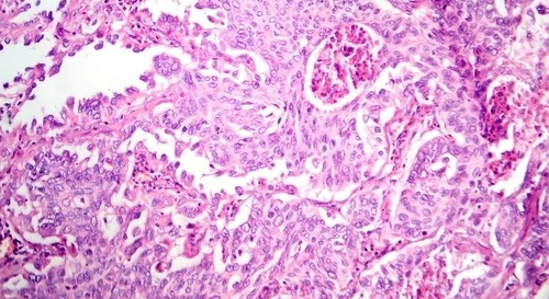
Colorectal cancer is diagnosed in an estimated 1.9 million people worldwide each year, often without symptoms in its early stages. Timely detection of precancerous lesions is therefore important, yet some can be difficult to distinguish on biopsy slides. Sessile serrated adenomas (SSAs), for example, may resemble benign hyperplastic polyps, complicating pathologic assessment. New findings suggest that artificial intelligence models may improve this distinction, helping pathologists identify these challenging lesions more accurately.
Researchers at Northeastern University’s Amal Lab for Precision Medicine (Boston, MA, USA) evaluated vision transformer models for histopathological classification of colorectal polyps. Unlike convolutional neural networks (CNNs), which typically analyze image features more locally, vision transformers can assess relationships across broader regions of an image, allowing a more global interpretation of tissue structure.
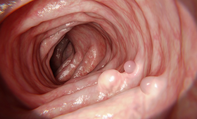
The study compared three vision transformer models with three CNN models using images of more than 3,000 colorectal polyps from a public dataset provided by the Department of Pathology and Laboratory Medicine at Dartmouth-Hitchcock Medical Center. The dataset included 1,545 benign hyperplastic polyps and 630 precancerous sessile serrated adenomas (SSAs). The best-performing vision transformer achieved 70% sensitivity for identifying precancerous lesions, compared with 40% for the top-performing CNN, while overall accuracy reached 86% versus 80%, respectively.
The findings were published in Lecture Notes in Computer Science under the title “Transformer Architectures Improve Minority-Class Detection in Histopathological Classification of Colorectal Polyps.” The researchers noted that the models were analyzed on one data set, and further testing on multiple data sets is required to validate the findings. The team has begun working directly with pathologists and gastroenterologists to test the systems in clinical workflows.
“Pathologists help verify the AI's findings through tissue examination, while gastroenterologists assess how those findings can guide patient care. Working with both groups in prospective studies allows us to evaluate these algorithms in real time, within clinical workflows, and determine how accurate, helpful, and useful they are in practice,” said Saeed Amal, Northeastern professor of bioengineering.
“Research like this, which rigorously identifies where AI succeeds and where it fails in imbalanced real-world data, is exactly the kind of foundational work needed to bring reliable AI screening tools into clinical practice and prevent more cancers before they start,” said Leon Toussaint, scientific manager at the National Foundation for Cancer Research.
Related Links
Amal Lab for Precision Medicine – Northeastern University