Label-Free Platform Combines Microfluidics and AI for Circulating Tumor Cell Analysis
Posted on 01 Oct 2026
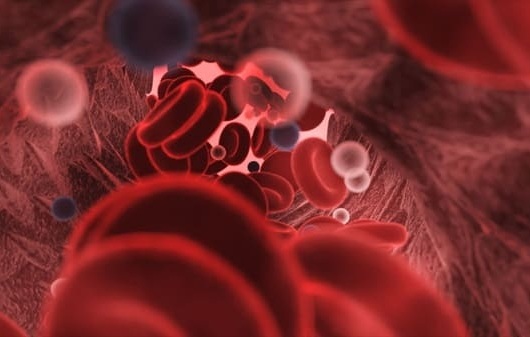
Liquid biopsy relies on detecting rare tumor-derived material in blood, but circulating tumor cells (CTCs) are especially difficult to capture because they are vastly outnumbered by normal blood cells. Despite their scarcity, CTCs can provide valuable information about cancer progression and treatment response, with concentrations sometimes as low as one to ten cells among a billion blood cells.
Researchers at Taiyuan University of Technology and Shanxi Bethune Hospital developed the platform for CTC analysis. The system integrates inertial microfluidic enrichment with YOLOv8-based deep learning for bright-field image recognition. The approach is designed to enrich tumor cells in complex blood backgrounds without relying on specific cell-surface markers.
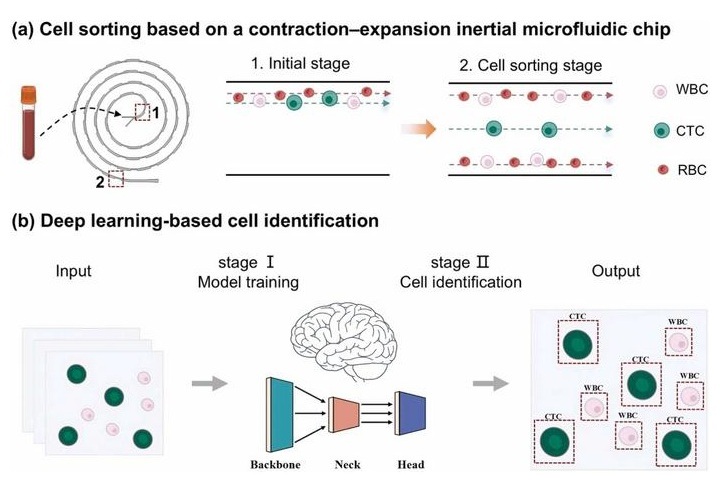
The microfluidic component uses a spiral chip containing engineered contraction-expansion structures. Because CTCs are generally larger than blood cells, different cell populations experience different forces as they move through the microchannel. The chip directs tumor cells and blood cells into separate flow paths, allowing tumor cells to be collected without antibody-based or marker-dependent capture.
The proof-of-concept study used artificial blood samples containing MCF-7 breast cancer cells and white blood cells. Tumor cells were enriched at the central outlet, while most blood cells were directed toward side outlets. The platform achieved an MCF-7 recovery rate of 89.2 ± 3.1% and a white blood cell removal rate of 86.9 ± 1.4%. The proportion of tumor cells increased from 9% before separation to 38% after enrichment.
For cell identification, the team used a YOLOv8 deep-learning model to recognize tumor cells from bright-field microscope images. Fluorescence images were used during model development to support annotation, but after training the model performed recognition using bright-field images alone. The model achieved 96.0% accuracy, 94.6% precision and recall, and 95.4% specificity in distinguishing tumor cells from non-tumor cells.
By combining microfluidic cell separation with AI-based image analysis, the platform targets two key challenges in CTC testing: isolating extremely rare tumor cells from complex blood samples and confirming their identity after enrichment. Because the workflow does not depend on labels or surface markers, it could streamline sample preparation while preserving cells for downstream uses such as single-cell sequencing and drug susceptibility testing. The findings also highlight the potential of pairing microfluidics with deep learning to expand rare-cell analysis.
The study was published in Biomedical Analysis under the title “Label-free Enrichment and Identification of Circulating Tumor Cells Integrating Inertial Microfluidics and Deep Learning.” The researchers described the work as a proof-of-concept study validated with MCF-7 cell lines and artificial blood samples rather than patient-derived specimens. The enrichment and recognition modules have also not yet been fully integrated into a single automated system. Future work will focus on additional tumor types, patient samples, system integration, and automation.
“The challenge of circulating tumor cell analysis is not only finding these rare cells, but also identifying them accurately after separation,” said Xiaochun Li, corresponding author from Taiyuan University of Technology. “By combining microfluidics with artificial intelligence, we hope to provide a simpler and more flexible approach for label-free CTC analysis and future downstream applications.”
Related Links
Taiyuan University of Technology