AI Matches Pathologists in Predicting Breast Cancer Prognosis from Immune Cells
Posted on 27 Aug 2026
Breast cancer is the most common cancer in Australian women, with more than 20,000 cases each year. Prognosis can be informed by counting tumor-infiltrating lymphocytes (TILs) on routine pathology slides, but assessments typically require expert review. In some settings, routine or widespread pathologist evaluation is not available. New findings demonstrate that AI can match human pathologists in using TILs to predict outcomes across common breast cancer subtypes.
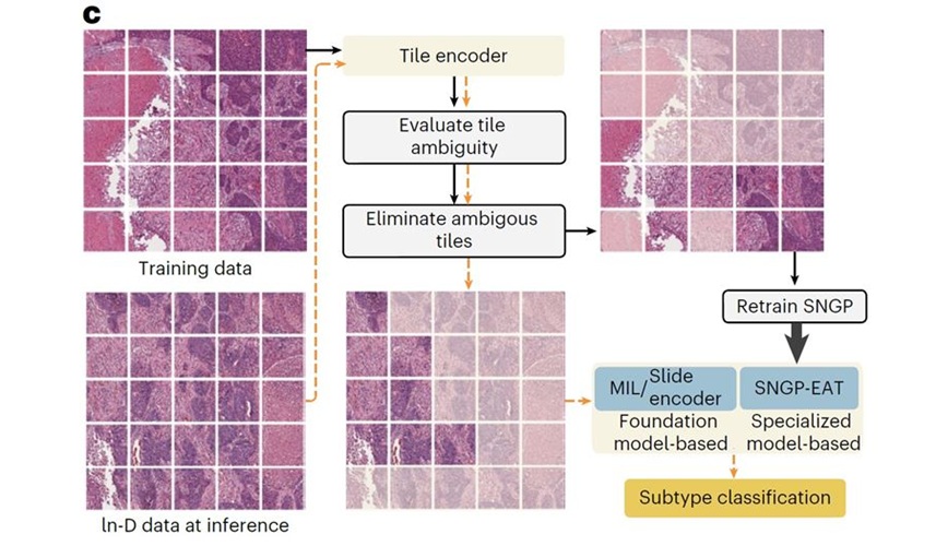
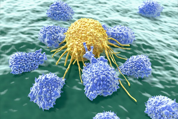
Peter MacCallum Cancer Centre (Peter Mac; Victoria, Australia) investigators evaluated AI models that quantify tumor-infiltrating lymphocytes (TILs) on routine breast tissue slides. TILs are immune cells whose higher levels reflect a stronger antitumor response and have been associated with improved outcomes across several breast cancer types. The researchers compared AI-derived TIL measurements with expert pathologist scoring and digital image analysis to determine whether the different approaches provided similar prognostic information.
One analysis, CATALINA, aggregated seven clinical trials in triple-negative breast cancer, with TILs from more than 1,300 patients scored by both a pathologist and an AI model. A second analysis examined more than 4,300 tumor samples from the Phase III APHINITY trial in early HER2-positive disease. Both studies were led by Peter Mac and were published simultaneously in The Lancet Oncology in 2026.
In the triple-negative cohort, AI and pathologist scores were not identical, but both methods similarly stratified prognosis, with higher TIL levels associated with better outcomes. In the HER2-positive cohort, expert scoring, digital image analysis, and AI approaches all showed that patients with higher TIL levels had improved outcomes. Patients with the highest TIL levels, particularly those with node-positive disease, appeared to benefit more when pertuzumab was added to standard care. AI also identified spatial immune-cell “hotspots” that provided prognostic information beyond simple cell counts.
The findings strengthen the case for TILs as a practical biomarker and indicate that AI could enable standardized assessment at scale, including in settings where expert pathology may be limited. Together, the studies encompassed more than 5,600 patients across two major forms of breast cancer. They conclude that AI models that count TILs should be more widely used, particularly where routine or widespread pathologist assessment is unavailable.
“Taken together, these two studies provide strong evidence that the immune response visible on a routine breast cancer pathology slide contains clinically important information. What is particularly reassuring is that we see this across two very different types of breast cancer and across thousands of patients treated in randomized clinical trials,” said Professor Sherene Loi, Peter MacCallum Cancer Centre.
“Pathologist assessment of TILs remains highly reproducible when performed using standardized methods, but AI gives us the opportunity to measure this biomarker objectively and at enormous scale, including in settings where expert pathology assessment may not be readily available. AI may also allow us to extract information that the human eye cannot readily quantify, such as the spatial organization of immune cells within a tumor. Ultimately, combining pathological and computational approaches may give us more information than either approach alone,” said Prof. Loi.
Related Links
Peter MacCallum Cancer Centre