New Review Highlights Intelligent Agents as Next Step for Digital Pathology
Posted on 24 Aug 2026
Digital pathology remains constrained by models that classify single images without mirroring how clinicians interrogate multiple slides, adjust magnification, and synthesize ancillary tests. This gap can hinder efficient case review and transparent reasoning, especially in complex oncology or rare disease workups. A growing need exists for systems that integrate evidence, represent uncertainty, and revise decisions across the diagnostic workflow. A new review describes intelligent agent architectures designed to align computational pathology with real-world practice.
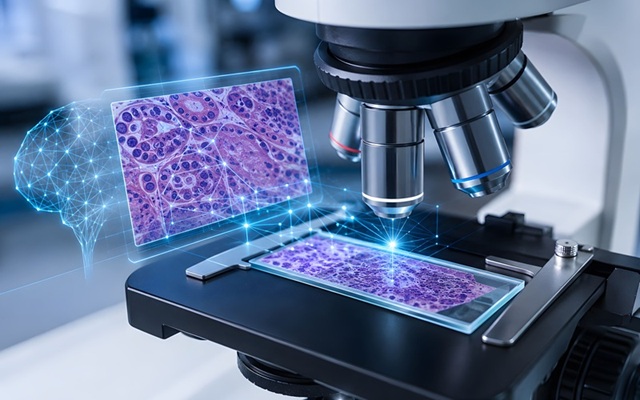
Researchers at the Department of Pathology, Peking Union Medical College Hospital, Chinese Academy of Medical Sciences and Peking Union Medical College, examine how “intelligent agents” could be organized to support computational pathology. The review was published online July 7, 2026, in the Medical Journal of Peking Union Medical College Hospital. The approach coordinates large language models (LLMs), vision-language models (VLMs), and specialized tools to mirror pathologists’ stepwise reasoning and documentation.

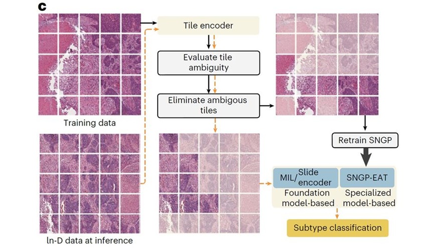
According to the review, the agent workflow spans three interconnected stages: low-magnification slide overview, diagnostic reasoning, and report generation, linked through memory and dynamic revision. During the overview stage, whole-slide images (WSIs) are navigated through a sequence of decisions about where to move, when to zoom, and when to stop, rather than analyzed through a single-pass prediction. This approach differs from conventional patch-based pipelines such as multiple instance learning (MIL), which can highlight relevant regions but do not explicitly model the search process.
During diagnostic reasoning, visual models extract morphological features while large language models (LLMs) organize higher-level diagnostic logic. Chain-of-thought (CoT) strategies structure observations, supporting evidence, and the exclusion of alternative diagnoses, while tool use allows the system to call dedicated image-analysis, statistical, or multimodal modules as needed. Retrieval-augmented generation (RAG) can further incorporate information from guidelines, textbooks, or annotated cases, potentially supporting evaluation of rare or atypical presentations.
At the reporting stage, emerging systems aim to integrate findings across multiple slides, retain salient information through memory mechanisms, retrieve similar historical cases, and adapt to pathologist feedback. However, the review identifies major barriers to clinical deployment, including incorrect memories, model drift, uncertain evidence tracing, privacy risks, and inconsistent outputs. Clinical usefulness will therefore depend on transparent reasoning, traceable evidence, controlled model updating, and rigorous evaluation in collaboration with practicing pathologists.
The authors emphasize that reliable end-to-end performance across real diagnostic workflows has not yet been demonstrated. Most published studies remain focused on visual question answering (VQA) or narrowly defined diagnostic tasks rather than integrated clinical pathways. The review therefore presents these systems as a roadmap toward potential benefits, including reducing time spent scanning large slides, organizing multimodal evidence, supporting differential diagnosis, and generating clearer reports, while calling for systematic trials to assess stability, reproducibility, data security, auditability, and clinical benefit.
Related Links
Peking Union Medical College Hospital