Ultrathin Metasurface Could Bring Quantitative Phase Imaging to Portable Devices
Posted on 18 Aug 2026
Point-of-care imaging often depends on bulky optical systems that have difficulty visualizing nearly transparent cells and tissues without staining. These limitations can slow diagnosis and restrict use at the bedside or in resource-limited settings. Although phase-based methods can reveal important morphological details, conventional systems often trade accuracy for speed and compactness. Researchers have now developed an ultrathin metasurface approach designed to capture quantitative phase information on smaller sensors, supporting more compact clinical imaging devices.
Researchers at the Australian Research Council Centre of Excellence for Transformative Meta-Optical Systems (TMOS), working across RMIT University and The University of Melbourne, have created a metasurface that supports quantitative phase microscopy (QPM). The device images microscopic objects that are nearly invisible to conventional intensity-based optics. It is designed to enable compact, field-deployable imaging for clinical and research settings.
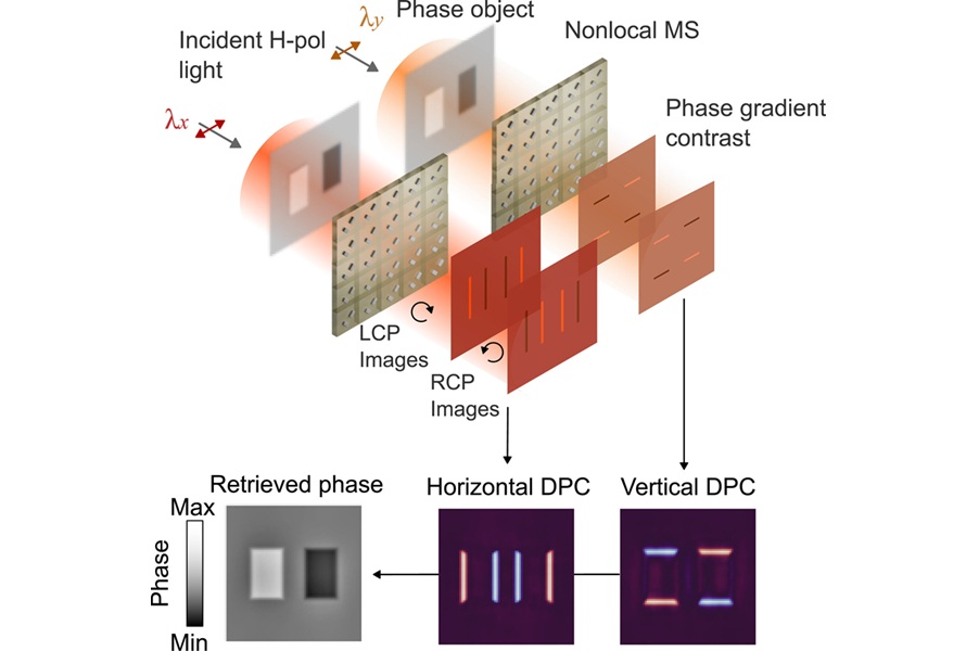
The metasurface is “nonlocal,” meaning it processes the entire image rather than redirecting individual rays like a lens. Because it can sit directly in front of the sample, it reduces space and alignment demands. It integrates multiple nanostructured elements onto a single ultrathin surface to deliver phase-contrast information with minimal hardware movement.
Quantitative phase microscopy (QPM) requires phase-gradient measurements along two perpendicular directions. Conventional approaches typically obtain these measurements by rotating optical elements or swapping components, adding time and complexity to the imaging workflow. The new design integrates two sets of wavelength-selective nanostructures onto a single surface, allowing users to switch between orthogonal measurements simply by changing the illumination wavelength. The researchers also incorporated differential phase contrast to reduce noise and improve reconstructed phase maps.
Published in Nanophotonics on July 1, 2026, the approach is intended to simplify imaging workflows for conditions in which cell shape and growth patterns are clinically relevant, including sickle cell disease, cancer, and neurodegenerative disorders. The researchers note that near-term challenges include the cost of supporting hardware and fabrication, as well as the need for precisely tuned dual-wavelength illumination. They are also exploring integration with commercial microscopes using an infrared light-emitting diode.
Related Links
TMOS