AI Links Organ-Specific Aging Patterns in Histology to Blood-Based Signals
Posted on 19 Aug 2026
Clinical decisions often rely on chronological age, yet organs can age biologically at different rates that remain hard to assess. Noninvasive detection of organ-specific aging could support disease monitoring and early diagnosis. Histology captures structural changes that reflect cumulative biological stress but are not easily discerned by clinicians. Researchers now demonstrate an AI approach that reads those patterns and links them to blood-based indicators of which organs are aging faster.
At the CeMM Research Center for Molecular Medicine of the Austrian Academy of Sciences, together with the Ludwig Boltzmann Institute for Network Medicine (LBI‑NetMed) at the University of Vienna, investigators developed artificial intelligence “tissue clocks” that estimate organ‑specific biological age from histological images. The models calculate organ‑level age gaps rather than a single whole‑body score. The approach is designed to detect tissue remodeling that is invisible to routine microscopic review.
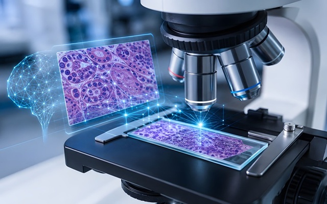
The method applies deep learning vision models to the microscopic architecture of high‑resolution images of tissue slices. Across 40 tissue types, age emerged as the dominant factor shaping tissue appearance, enabling construction of organ‑specific clocks. The image‑derived tissue age gaps were then linked to blood‑based gene‑expression profiles from the same individuals to build predictors that estimate tissue‑specific aging from blood samples.
The analysis drew on the Genotype‑Tissue Expression (GTEx) Project, which includes tissue samples from 983 individuals spanning 40 organs such as brain, heart, lung, pancreas, skin, and intestine. In total, 25,712 histology images—representing roughly 480 million image tiles—were processed with state‑of‑the‑art computer vision. This large‑scale design enabled robust training and evaluation of organ‑targeted aging models and their blood‑based counterparts.
The tissue clocks achieved a mean prediction error of 4.9 years and outperformed DNA‑based aging estimates in capturing tissue‑specific pathology. Predicted biological age correlated with hallmarks of aging, including telomere shortening, tissue pathology, and the number of chronic diseases. Organ trajectories varied: lung, kidney, pancreas, and adrenal gland exhibited accelerated aging between ages 20 and 40; the uterus showed a shift around menopause; kidney failure was linked to accelerated aging across multiple tissues, and diabetes produced pronounced effects in the pancreas.
Blood‑derived predictors recapitulated organ‑linked aging signals associated with Alzheimer’s disease, Crohn’s disease, cystic fibrosis, vasculitis, diabetes, and stroke. For example, the strongest signal in Alzheimer’s disease localized to brain, whereas Crohn’s disease reflected accelerated aging across the gastrointestinal tract. The authors state that such approaches could contribute to minimally invasive diagnostics for monitoring organ health and disease progression. The study, “Histological aging signatures for monitoring tissue-specific aging and disease,” was published in Nature Medicine on August 14, 2026.
“Our tissues carry a remarkably detailed record of the aging process. By combining histology images with artificial intelligence, we can detect patterns of biological aging that are invisible to the human eye and begin to understand how aging unfolds differently across the body,” said André Rendeiro, Principal Investigator at CeMM and corresponding author of the study.
“This study highlights that aging is not simply a matter of chronological time. Different organs age in different ways, and these processes appear to be shaped by both systemic and tissue-specific factors,” ,” said Yimin Zheng, the third co-first author of the study.
Related Links
CeMM Research Center for Molecular Medicine
LBI‑NetMed