AI Uses H&E Slides to Predict Key Biomarkers Across 32 Cancers
Posted on 15 Aug 2026
Molecular profiling is essential for characterizing solid tumors, but many laboratories still rely on separate genetic assays to detect alterations such as TP53 mutations. These workflows can be resource-intensive and may delay access to prognostic information across diverse cancer types. Pathologists need tools that extract molecular signals directly from routine stain preparations without exhaustive annotations. A new study shows that a single AI system can derive TP53 biomarkers, tumor classification, and survival indicators from standard hematoxylin and eosin images spanning 32 cancers.
At the Menzies Institute for Medical Research, University of Tasmania, investigators developed an AI-based Vision Transformer model that analyzes routine hematoxylin and eosin (H&E)-stained whole-slide images (WSIs) to produce multiple slide-level outputs. The system was designed to infer TP53 mutation status, estimate TP53 RNA expression, classify tumor type, and predict survival-related outcomes, including overall survival (OS) and progression-free interval (PFI). By linking histomorphology with genomic and clinical endpoints, the approach aims to streamline information retrieval from existing pathology materials.
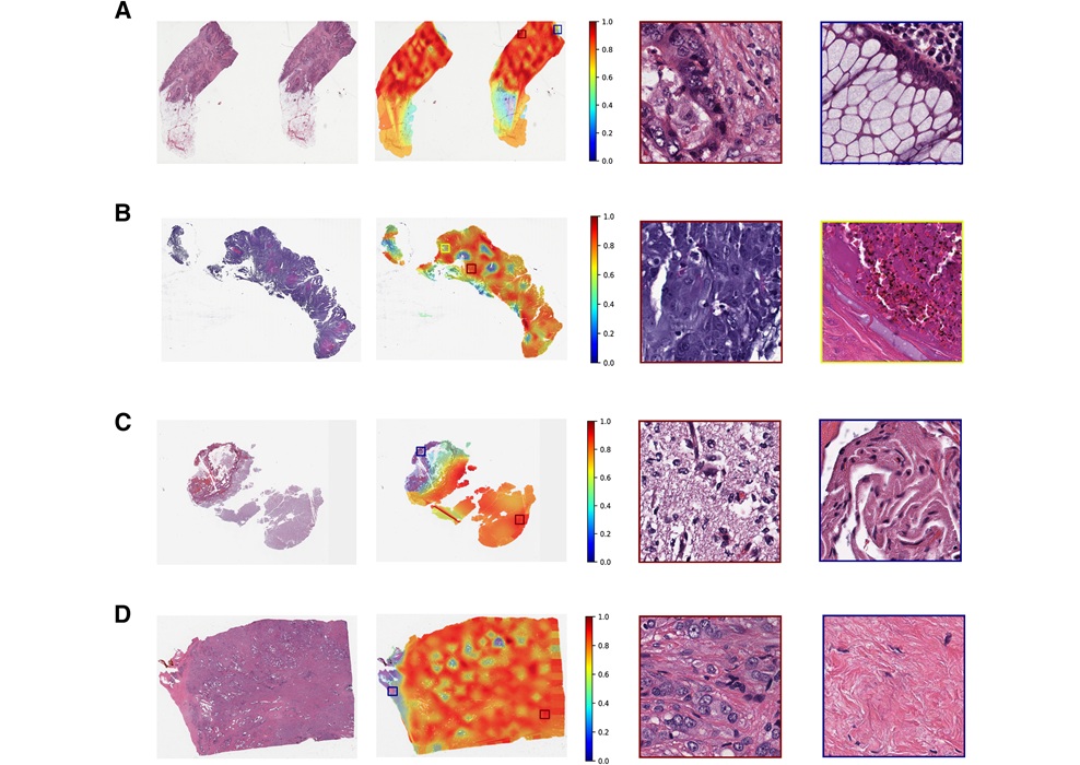
The model was trained using more than 11,000 primary tumor cases from the Pan-Cancer Atlas, each paired with somatic mutation, RNA-sequencing, and clinical outcome data. To address the scale and complexity of WSIs and the challenge of obtaining pixel- or region-level annotations, the team implemented a weakly supervised learning strategy. This enabled learning from slide-level labels to identify relevant morphological patterns across image patches without exhaustive expert markup.
In an independent validation set comprising 1,729 slides and covering 32 solid tumor types, the system achieved an area under the receiver operating characteristic curve (AUROC) of 0.766 for detecting TP53 mutations. Beyond mutation status, the model also inferred TP53 RNA expression levels and recovered tumor taxonomy directly from WSIs. These findings indicate that multi-task computational pathology can connect routine diagnostic imaging with molecular features at scale.
The study was published in The American Journal of Pathology. Affiliations represented in the work include the Menzies Institute for Medical Research and School of Medicine, University of Tasmania, and Pandani Solutions Pty Ltd. The authors note that comprehensive genomic testing remains costly or inaccessible in many settings, positioning the method as a complement to, rather than a replacement for, standard molecular assays.
“Standard molecular profiling for TP53 mutations is often costly and inaccessible in underprivileged or remote clinical settings. We wanted to develop a more practical tool for pathologists. Currently, most deep learning-based models are used for single-model concepts; one model for one task. We developed a single model that can generate seven outputs simultaneously from the whole-slide histopathology image, including TP53 mutation status, TP53 RNA expression, tumor type and survival-related outcomes at the slide level,” said Alex W. Hewitt, Ph.D., Menzies Institute for Medical Research and School of Medicine, University of Tasmania.
“This approach could help identify patients who may benefit from confirmatory molecular testing, support triage in settings with limited genomic testing, and provide additional decision support to clinicians. Importantly, this method should be viewed as complementary to molecular testing, not a replacement. Its potential impact is strongest as a screening, prioritization or decision-support tool within broader diagnostic pathways,” stated Abadh K. Chaurasia, Ph.D., Menzies Institute for Medical Research, University of Tasmania, and Pandani Solutions Pty Ltd.
Related Links
Menzies Institute for Medical Research