Molecular Imaging to Reduce Need for Melanoma Biopsies
Posted on 27 Feb 2026
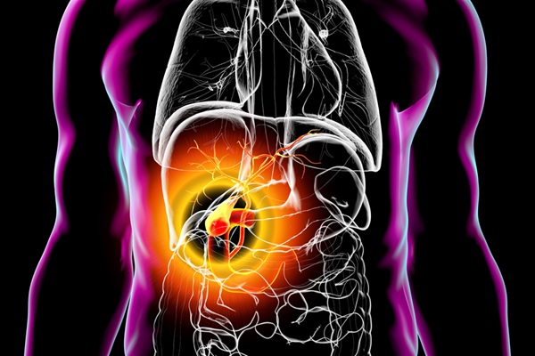
Melanoma is the deadliest form of skin cancer and accounts for the vast majority of skin cancer-related deaths. Because early melanomas can closely resemble benign moles, clinicians often rely on visual inspection and adopt a “when in doubt, cut it out” strategy, leading to many precautionary biopsies. Definitive diagnosis requires removing suspicious lesions, an invasive procedure that may be unnecessary in many cases. Now, a new noninvasive imaging technology has shown promise in identifying invasive melanoma by detecting molecular signals in suspicious skin lesions without removing tissue.
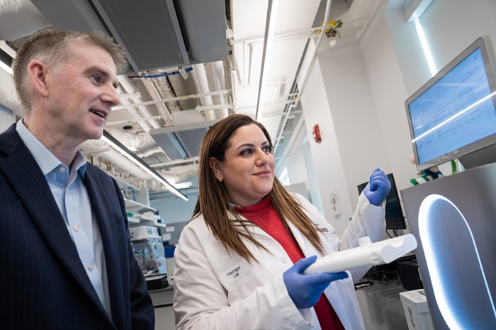
The system, known as Skin Fluorescent Imaging (SFI), has been developed by Orlucent, Inc. (Boston, MA, USA) and analyzes the molecular composition of moles and lesions using a targeted fluorescent dye and real-time image analysis. In a Phase 2 clinical trial conducted at Huntsman Cancer Institute at the University of Utah (Salt Lake City, UT, USA), in collaboration with Orlucent, clinicians evaluated the system by applying a fluorescent dye that binds to the αvβ3 protein, a marker associated with tumor growth and aggressiveness.
")
A handheld imaging device captured fluorescence signals from the lesion and surrounding tissue, and a machine-learning algorithm analyzed the data to determine the likelihood of melanoma. Researchers analyzed 240 pigmented lesions that had been flagged as suspicious during routine skin examinations. The system successfully identified all cases of invasive melanoma, the most aggressive form of the disease.
The study, published in the Journal of the American Academy of Dermatology International, also demonstrated that SFI could distinguish between normal moles and atypical moles that may have precancerous features. These results suggest the technology can improve screening precision while reducing unnecessary biopsies. By providing molecular-level information during a skin exam, SFI may help clinicians better determine which lesions truly require biopsy. This targeted approach could reduce invasive procedures while maintaining high sensitivity for dangerous melanomas. The technology may be particularly beneficial in regions such as the Mountain West, where melanoma rates are high and access to dermatologists can be limited in rural areas. Additional validation studies will be needed before widespread clinical implementation.
“By identifying the molecular signals that drive melanoma early on, SFI has the potential to help clinicians decide which suspicious lesions to biopsy while sparing patients unnecessary biopsies,” said co-leader Douglas Grossman, MD, PhD. “SFI is a promising step forward in noninvasive approaches to catching melanoma early and reducing biopsies of moles.”
Related Links:
Huntsman Cancer Institute
Orlucent, Inc.