AI-Based Digital Pathology Platform Improves Lung Cancer Diagnosis
Posted on 26 Aug 2024
Lung cancer ranks among the most prevalent and fatal cancers worldwide. Current treatment strategies for lung cancer patients rely on pathological examinations, which can reveal genetic mutations specific to the patient's cancer, facilitating personalized treatment approaches. Over the last few years, pathology has evolved dramatically due to digital advancements, making traditional microscopes obsolete. Tissue samples are now digitized and analyzed via computer screens, which is essential for employing sophisticated artificial intelligence (AI)-based analytical methods. These AI technologies can extract additional insights from pathological tissue sections that were previously unattainable.
A team of researchers at the University of Cologne (Cologne, Germany) has developed an AI-driven digital pathology platform that can revolutionize the analysis of lung cancer tissues. This platform utilizes newly developed algorithms to perform fully automated examination of digitized lung cancer tissue sections, offering faster and more precise analyses than traditional methods. Detailed in their publication in the journal Cell Reports Medicine, the platform is founded on the most extensive and high-quality dataset available, enabling it to process H&E-stained whole-slide images (WSIs) of both resection and biopsy specimens from non-small cell lung cancer (NSCLC) patients. It can accurately segment all relevant tumor and benign tissue classes at the pixel level.
")
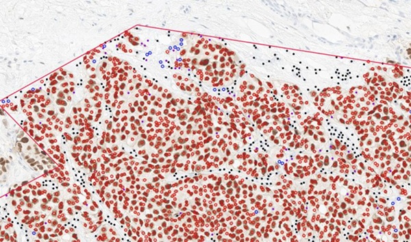
The core of this platform is a cutting-edge multi-class tissue segmentation algorithm, marking a significant leap in the accuracy and precision of lung cancer pathology. This development is among a handful of published studies that focus on tissue detection and classification in WSIs from lung cancer patients. Furthermore, the research team has identified four prognostic parameters that have proven effective in stratifying NSCLC patients prognostically, which could be crucial for determining patient eligibility for adjuvant therapy following surgery. To support ongoing academic research and enhance algorithm development and benchmarking, the team has made four of their annotated test datasets publicly available. This innovative platform is poised to transform various diagnostic, prognostic, and predictive applications within the field.
“We also show how the platform could be used to develop new clinical tools,” said physician Dr. Yuri Tolkach from the Institute of General Pathology and Pathological Anatomy at University Hospital Cologne, who led the study. “The new tools can not only improve the quality of diagnosis, but also provide new types of information about the patient’s disease, such as how the patient is responding to treatment.”
Related Links:
University of Cologne
 Assay.jpg)